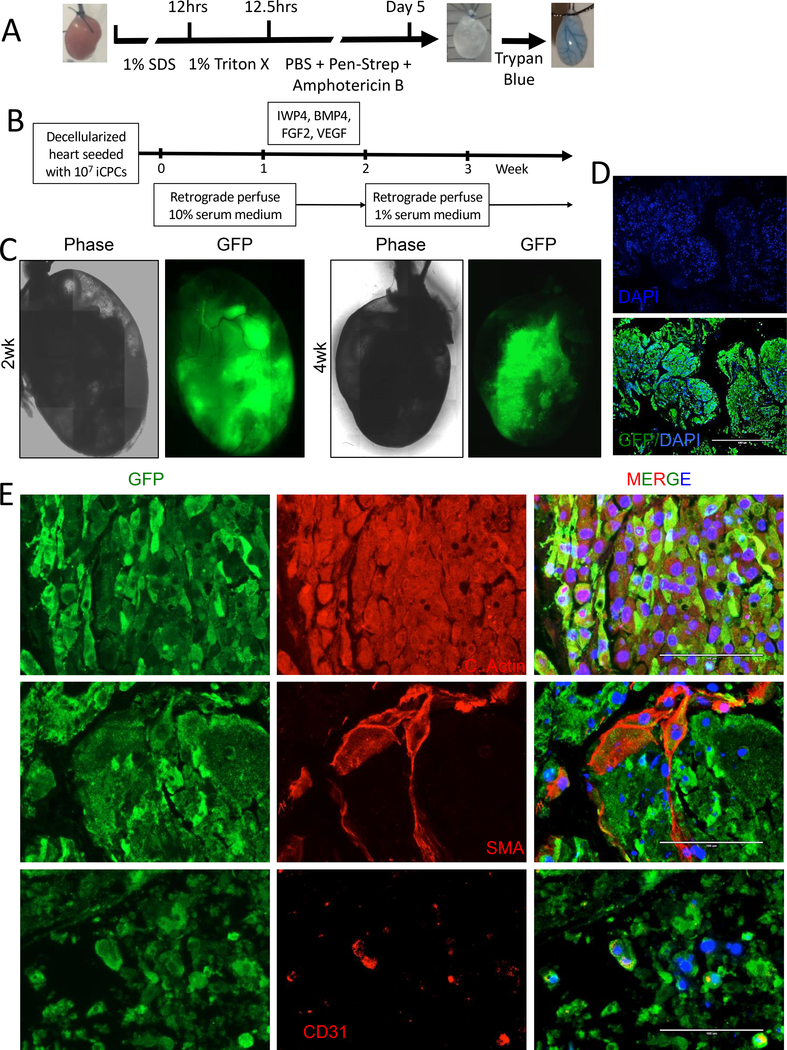
Fig. 3. iCPCs repopulate decellularized heart scaffold and differentiate into cardiac lineage cells.
(A) Schematic protocol for decellularization with images of aorta cannulated mouse heart before, after decellularization, and after decellularization with trypan blue perfusion. (B) Schematic of iCPC repopulation with 10 million GFP labelled iCPCs injected retrograde via an aortic cannula into decellularized whole heart scaffold. Scaffolds were placed in the incubator for 1–3 hr to allow cell attachment and then perfused for 4–5 weeks with media and growth factors/small molecules as indicated. (C) Live imaging was performed at various time points and showed that iCPCs attach and repopulate the decellularized scaffolds with phase contrast and epifluorescence for GFP. (D) GFP staining 3 weeks after recellularization revealed that iCPCs migrated and colonized the left ventricular myocardium region of the decellularized heart scaffold. (E) Epifluorescence images of sections from 4 week recellularized scaffolds for GFP marking iCPC progeny, DAPI to label nuclear DNA, cardiac actin for cardiomyocytes, smooth muscle actin from smooth muscle, and CD31 for endothelial cells. Scale bar = 1000uM for D and 100uM for E.

