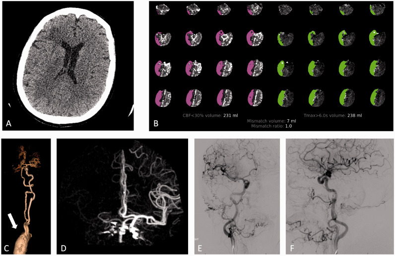
Figure 3.
Case 2. NCCT (a) demonstrates subcortical hypoattenuation in the right frontal lobe. CTP (b) demonstrates large area of decreased CBF and prolonged TMax within the right MCA territory and right posterior fossa. CTA 3D reconstruction of the great vessels (c) and volume rendered reconstruction of the cerebral vasculature (d) demonstrate non-opacification of the brachiocephalic artery (arrow), right CC artery, right SA, right VA and right MCA. Anteroposterior (e) and lateral (f) angiography images following right carotid artery injection demonstrate opacification of the right carotid, right MCA and right ACAs without large vessel occlusion.

