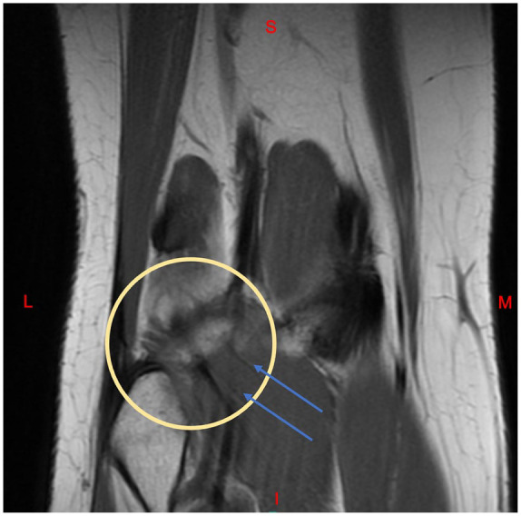
Figure 3.

Coronal MRI of the right knee. The distal popliteal muscle is visualized (blue arrows), however a gap is seen where the tendon should attach proximally at the fibular head and lateral femoral condyle (yellow circle). A few proximal tendinous fibers remain.
Abbreviations: S, Superior; I, Inferior; M, Medial; L, Lateral.
