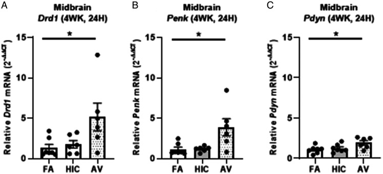
Figure 9.
Four-week Aspergillus versicolor exposure elevates basal ganglia neurotransmission genes in the midbrain. Eight week-old female B6C3F1/N mice were exposed in a nose only chamber to filtered air (FA), 3 × 10⁵ spores of heat-inactivated Aspergillus versicolor conidia (HIC), or 3 × 10⁵ spores of viable Aspergillus versicolor (AV) twice weekly, for 4 weeks and markers of neuroinflammation were assessed in the midbrain 48H after the last exposure. Relative Drd1 (A), Penk (B), and PDyn (C) mRNA levels in the midbrain were determined using qRT-PCR. Individual data points for an experimental animal are represented as black dots. Values were normalized to Gapdh using the 2-ΔΔCT method and are the mean ± SEM. *p<.05 vs. filtered air control; n = 6–7.

