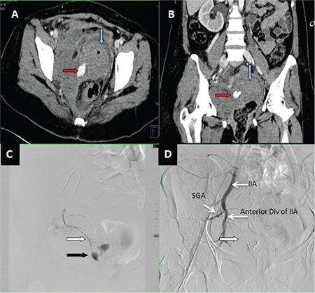
Figure 2.

CT angiography images (A, B) showing pseudoaneurysm (red arrow) and surrounding hematoma (blue arrow) in the right lateral wall of the vagina. DSA spot images (C, D) of the same patient showing pseudoaneurysm (black arrow) arising from the right vaginal artery (white arrow), which was embolized with a 30% glue injection, and post embolization angiogram (D) showed non-filling of pseudoaneurysm suggestive of successful embolization
IIA: Internal iliac artery, SGA: Superior gluteal artery, CT: Computed tomography
