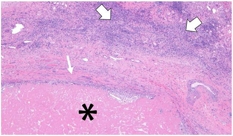
Fig. 2.

A slide showing part of a right hepatectomy specimen containing a 5.0 cm necrotic mass with no viable tumor. The Hematoxylin & Eosin stain at × 10 magnification shows the necrotic mass (*) rimmed by fibrous scar (small arrow) with peri-tumoral inflammatory cells (large arrows)
