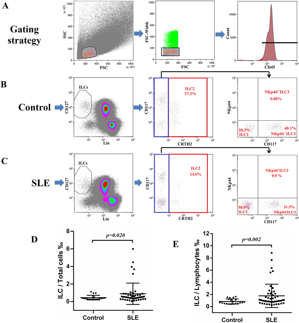
Fig. 1.
Increased frequency of ILCs in peripheral blood of SLE patients. PBMCs from healthy controls and SLE patients were stained for flow cytometry. (A) Single CD45+ lymphocytes were selected for ILCs analysis. ILCs were defined as Lin−CD127+ cells. Type 1, 2 and 3 ILC are identified by the expression of CRTH2, CD117 and NKp44. Representative gating strategy of ILCs in a healthy control (B) and a SLE patient (C). Percentages of ILC/Total cells (PBMC) (D) and ILC/Lymphocytes (E) in SLE patients (N = 49) and healthy controls (N = 20).

