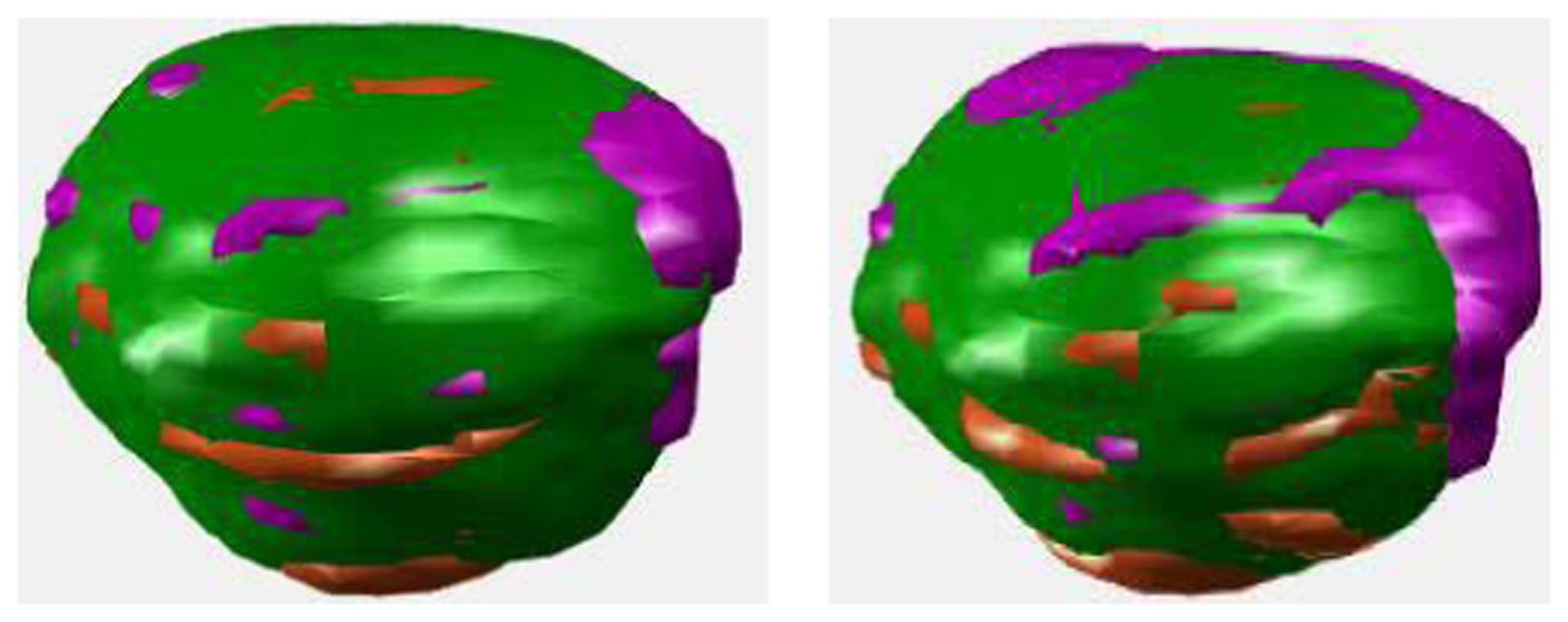
Fig. 3.

Tumor volumes segmentated in PET (first column) and CT (second column), where, as compared to the ground truth, where, the green region consists of the true positive and true negative voxels, the magenta region consists of the false positive voxels, while the orange region consists of the false negative voxels.
