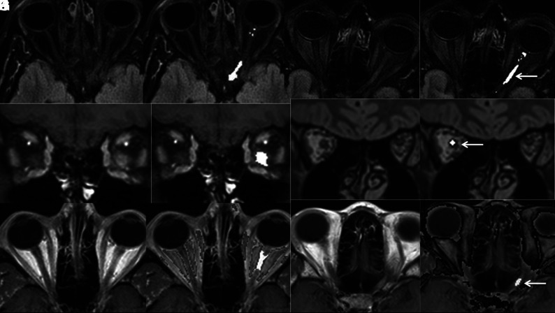
FIG 1.
Axial FLAIR (A and B), coronal CE-T1WI (C and D), and axial CE-T1WI (E and F) images from routine brain MR imaging of a patient with clinically proven left optic neuritis before (A, C, and E) and after (B, D, and F) processing with a proprietary postprocessing algorithm showing accentuation of the signal intensity of the diseased optic nerve and the unaffected contralateral optic nerve. Axial FLAIR (G and H), coronal 3D-FLAIR (I and J), and axial CE-T1WI of different patients with clinically proven unilateral optic neuritis again demonstrating accentuation of intensity in diseased optic nerves (arrows) after processing (H, J, and L). Note that the detection of alteration in the optic nerve signal is especially challenging in images obtained without fat saturation (E, I, and K).

