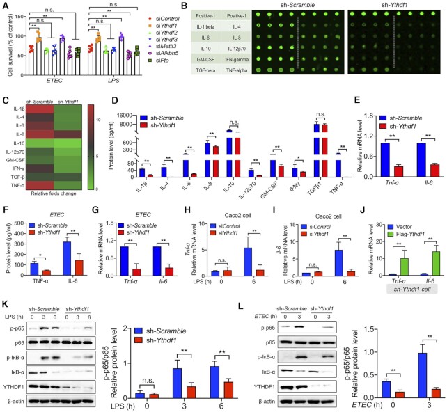
Figure 1.
YTHDF1 mediates the bacterial immune response in Intestinal epithelial cells. (A) Summary of the survival of IPEC-J2 cells transfected with a control siRNA or Ythdf1, Ythdf2, Ythdf3, Mettl3, Alkbh5, Fto siRNA and then either infected with ETEC (left) or stimulated with LPS (right). (B) Laser-scanning map of a porcine cytokine antibody array in LPS-treated cells expressing either a scrambled shRNA or the Ythdf1 shRNA. Each cytokine was measured in quadruplicate, and the table at the left indicates the cytokines. The top row is a positive control. (C) Heat map showing the fold change in signal intensity measured in (B), relative to cells expressing the scrambled shRNA. (D) The indicated cytokines were measured in the supernatants of IPEC-J2 cells expressing either the scrambled shRNA or Ythdf1 shRNA, 6 h after LPS stimulation. (E) Summary of Tnf-α and Il-6 mRNA measured 6 h after LPS stimulation in IPEC-J2 cells expressing either the scrambled shRNA or Ythdf1 shRNA, expressed relative to Gapdh mRNA. (F, G) Summary of TNF-α and IL-6 protein in the supernatant and Tnf-α and Il-6 mRNA levels (expressed relative to Gapdh mRNA) in ETEC-infected IPEC-J2 cells. (H, I) Summary of Tnf-α (H) and Il-6 (I) mRNA measured in Caco2 cells 6 h after LPS stimulation, expressed relative to Gapdh mRNA. (J) Summary of Tnf-α and Il-6 mRNA levels in IPEC-J2 cells co-expressing the Ythdf1 shRNA and an empty vector or FLAG-Ythdf1, expressed relative to Gapdh mRNA. (K, L) Western blot analysis and summary of the indicated proteins measured in IPEC-J2 cells expressing either the scrambled shRNA or Ythdf1 shRNA before and after LPS stimulation (K) or ETEC infection (L).

