Figure 1.
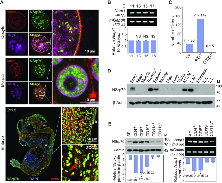
NSrp70 is preferentially expressed in early embryonic tissues and in lymphoid cells. (A) Immunohistochemistry for NSrp70. Oocyte (top), morula (middle), and fetal embryo E11.5 (bottom) were isolated from WT mice, stained with anti-NSrp70 antibody followed by FITC-conjugated 2nd antibody (green), TRITC-phalloidin (red), and DAPI (magenta and blue), and then visualized by confocal microscopy. (B) Fetal embryos from gestation day 11.5, 13.5, 15.5 and 17.5 mice were isolated. Nsrp1 mRNA was detected by RT-PCR (blots) and density was represented by bar graph. mGapdh was used as a loading control. E, embryonic day; bp, base-pair. (C) Intercrossing the heterozygous Nsrp1+/GT mouse produced no offspring homozygous for the allele containing the gene-trap vector. GT, gene-trap vector; +/+, wild-type; +/GT, heterozygous Nsrp1+/GT; GT/GT, homozygous Nsrp1GT/GT. (D) Tissue distribution of NSrp70 was determined by western blot analysis in 8-week old mice. M.L.N, mesenchymal lymph node; L.N., lymph node; S.I., small intestine; L.I., large intestine; M, molecular mass (KDa). (E) Western blot (left) and RT-PCR (right) for NSrp70 in mouse immune cells. β-actin and mGapdh were shown as loading controls. SP, splenocytes. All data shown are representative of three independent experiments. The bar graphs indicate the mean ± standard deviation of the indicated protein blot or RNA gel densitometry presented with respect to β-actin or mGapdh (B and E).

