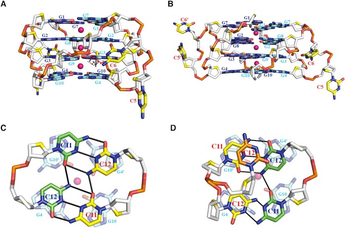
Figure 5.
Detailed conformations of cytosines in the tetrameric G-quadruplex formed by d(G4C2)2 in K+ (magenta sphere). The conformation of propeller loop, C5 and C6, in each tetrameric G-quadruplex of (A) Form-1/7 and (B) Form-1/1. (C) The conformation of the C11 and C12 bases of chain C forming Form-1/1 located at the 3′- end in the unit cell. (D) The conformation of the C11 and C12 bases of chain A (green) forming Form-1/1 and B (yellow) forming Form-1/7. Another conformation observed for the C12 base of chain B is colored by orange. The hydrogen bonds are represented by red dash and solid black lines.

