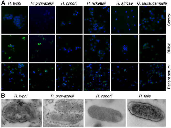
Fig 1. BNI52 specifically detects TG rickettsiae.
(A) L929 cells were infected with the indicated TG and SFG rickettsiae or Orientia tsutsugamushi and stained with isotype antibody as a negative control (upper panel), BNI52 (middle panel) or serum from patients suffering from the respective infection (lower panel). Bacteria are shown in green. Nuclei were stained with DAPI (blue). (B) L929 cells were infected with TG rickettsiae (R. typhi, R. prowazekii), SFG rickettsiae (R. conorii) or transitional rickettsiae (R. felis), stained with BNI52 and gold-labeled secondary antibody and analyzed by electron microscopy. The antibody binds to the TG rickettsiae R. typhi and R. prowazekii but not to the SFG rickettsiae R. conorii, R. rickettsii and R. africae or to R. felis.

