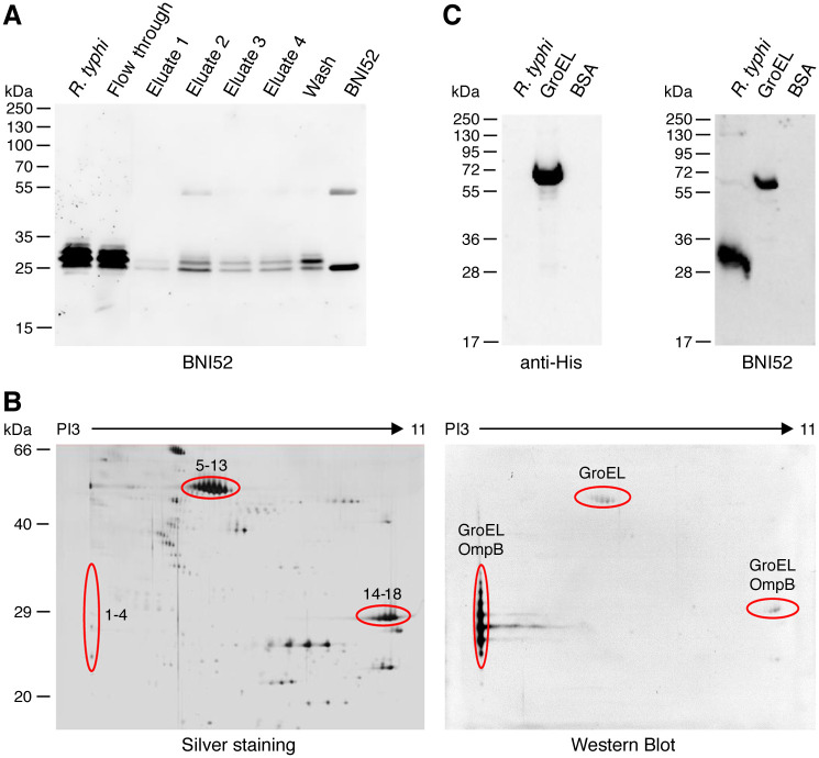
Fig 5. BNI52 binds the GroEL protein of R. typhi.
(A) Proteins in the lysate of R. typhi were precipitated with the BNI52 antibody by a protein A/G column. A Western Blot was performed from total lysate (R. typhi), flow through, eluates and wash fraction. BNI52 antibody was loaded as a control. The membrane was incubated with the BNI52 antibody. The antibody predominantly precipitates a 30 kDa protein. (B) For the identification of the antigen lysate of R. typhi was separated by two-dimensional SDS PAGE. Gels were silver stained (left) and applied to Western blotting. The membrane was incubated with the BNI52 antibody (right). The indicated protein spots (1–4, 5–13 and 14–18) of the silver stained gels that were recognized by the antibody in Western blots were analyzed by mass spectrometry. These analyses revealed the presence of GroEL in all spots recognized by BNI52. In addition, OmpB peptides were found in the lower molecular weight spots 1–4 and 14–18 but not in the 60 kDa spots. (C) His-tagged GroEL protein of R. typhi was expressed in E. coli, purified and subjected to Western blotting. Total lysate from R. typhi and BSA protein were used as a control. The membranes were incubated with a polyhistidine antibody (left) or the antibody BNI52 (right). The BNI52 antibody recognizes the 60 kDa recombinant GroEL protein but only weakly binds to the 60 kDa protein in R. typhi lysate.

