. 2021 Jun 1;10:e67789. doi: 10.7554/eLife.67789

Figure 5. SsaP is secreted during HeLa cell infection.

(A) SPIEDAC labeling of secreted SifA-Y65TCO with fluorogenic SiR-Tz. HeLa cells were infected with the ssaP null mutant of Salmonella expressing SsaP-Y65TCO in the presence of TCO*Lys for 12 hr. After 12 hr post infection, HeLa cells were incubated with 1.5 μM SiR-Tz in DMEM with 10% FBS growth media for another 2 hr, followed by extenstive wash out of excess dye with fresh growth media as described in Materials and methods. At 16 hr post-infection, cells were fixed. Cells were also immuno-stained for the endosomal membrane marker LAMP1 (green), LPS (Red) and DAPI (Blue) and imaged by spinning-disk SIM. From the merged image (right), it is evident that SsaP is present within the LAMP1-positive endosomes. A higher magnification spinning-disk SIM image of a SIF-positive HeLa cell from the boxed region (i) is shown on the right-most panel without the green channel displayed (i). It clearly shows secreted SsaP from Salmonella (boxed region). (B) SsaP was absent in infected cells that lacked ssaP. HeLa cells that were infected with an ssaP null mutant in the presence of TOC*Lys lack an SsaP signal. Note the absence of SIFs in the merged image (right) in the ssaP null strain. The data are representative of at least three independent experiments. Scale bar, 5 µm (A and i) and 10 µm (B).

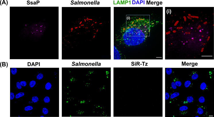
Figure 5.

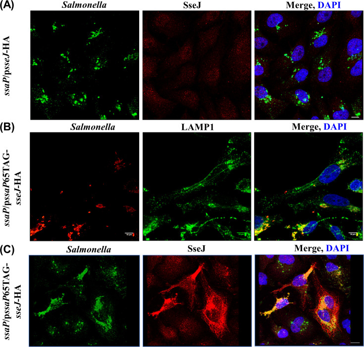
Figure 5—figure supplement 1. An ssaP null mutant is defective in SPI-2 secretion.

Figure 5—figure supplement 1.

(A) HeLa cells were infected with the ssaP null mutant for 16 hr, fixed and immuno-stained for LPS (green), SseJ (red) and DAPI (blue). In the absence of ssaP, SseJ is not secreted. (B) TCO*Lys-incorporated SsaP was functional. HeLa cells were infected with Salmonella complemented with pssaP65TAG-sseJ-HA, fixed and immunostained for LPS (red), LAMP1 (green) and DAPI (blue). SIF formation was observed in cells infected with Salmonella complemented with pssaP65TAG-sseJ-HA. (C) Engineered SsaP rescued SPI-2 secretion. HeLa cells were infected with Salmonella complemented with pssaP65TAG-sseJ-HA, fixed and immunostained for LPS (Green), SseJ (red), and DAPI (blue). SseJ (red) was secreted in the complemented strain, but not in the ssaP null strain (n = >10, independent experiments). Scale bar, 10 µm.
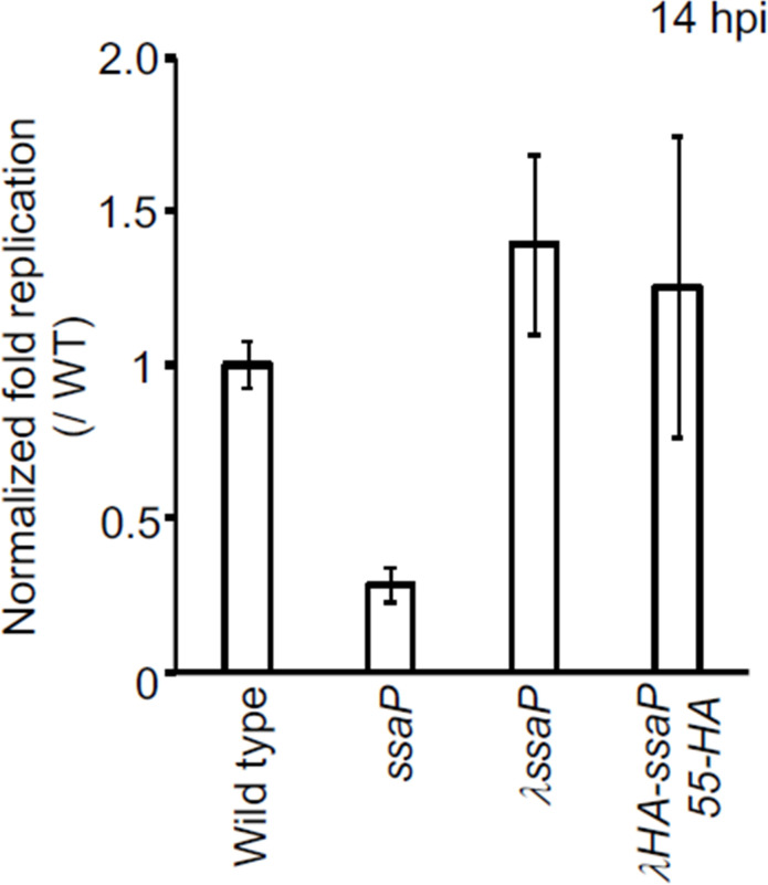
Figure 5—figure supplement 2. Survival of S. Typhimurium SsaP-55HA mutant in HeLa cells.

Figure 5—figure supplement 2.

Strains of Salmonella wild-type, ssaP null, ssaP/λssaP (complemented with ssaP integrated a λatt), and ssaP/ λssaP-55HA (ssaP with an HA tag at amino acid 55) were grown in LB to late exponential phase and added to HeLa cells at an MOI of 100:1 for 30 min. At 2 hr and 14 hr, cells were lysed and the intracellular bacteria were enumerated. The normalized fold changes were relative to those of the wildtype (set to 1). The error bars represent the mean ± standard deviation (n = 3).
Figure 5—figure supplement 3. SsaP is secreted.

Figure 5—figure supplement 3.

Strains of Salmonella containing ssaP/λssaP-55HA (left) ssaP/λssaP-110HA (middle and right) were grown in LB to late exponential phase and added to HeLa cells at an MOI of 100:1 for 30 min. The cells were fixed and immuno-stained for SsaP (red). A representative image at 14 hr post infection is shown. DAPI-stained DNA is in blue and GFP-expressing Salmonella are shown in green. The middle and right panels are of SsaP at different z positions.
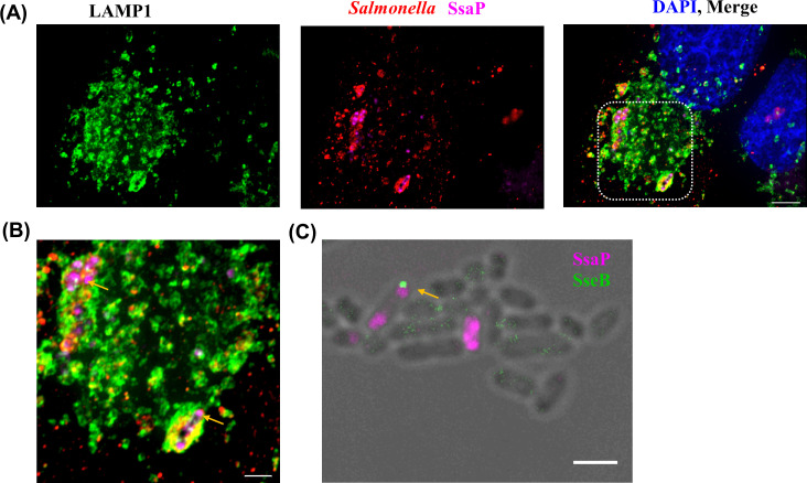
Figure 5—figure supplement 4. SsaP was associated with bacteria and mostly localized to the poles during early infection times.

Figure 5—figure supplement 4.

(A) HeLa cells were infected with an ssaP null mutant of Salmonella expressing SsaP-YTCO*Lys in the presence of TCO*Lys. After 6 hr post infection, HeLa cells were incubated with 1.5 μM SiR-Tz in DMEM with 10% FBS for another 1 hr, followed by extenstive washing of excess dye with fresh growth media. At 8 hr post-infection, cells were fixed and immunostained for the endosomal membrane marker LAMP1 (green, left panel), LPS (red) and DAPI (blue). Scale bar, 5 µm. (B) Zoomed view of inset. Yellow arrow indicates polarly localized SsaP. Images were acquired on a spinning-disk SIM, as described in Materials and methods. Scale bar, 2 µm. (C) Not every pole where SsaP was located had an injectisome. Salmonella cells expressing SsaP-Y65TCO in the presence of TCO*Lys were labeled with SiR-Tz as described in Materials and methods. The same cells were immuno-stained for SseB using rabbit anti-SseB antibody (green) and imaged along with SiR fluorescence (magenta). The arrows indicate cells that have both SsaP and SseB. The data are representative of at least three independent experiments. Scale bar, 2 µm.
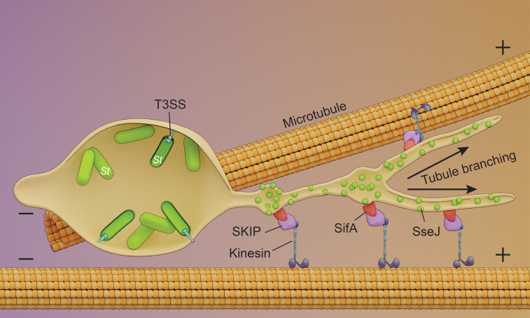
Figure 5—figure supplement 5. Schematic of endosomal tubule (SIF) formation.

Figure 5—figure supplement 5.

Upon entry into the host, Salmonella resides within an acidic vacuole. The histidine kinase EnvZ senses the acid pH of the Salmonella cytoplasm (Park et al., 2017) and drives OmpR-dependent up-regulation of the SsrA/B two-component system upon acidification. SPI-2 T3SS secreted effectors SseJ and SifA are present on the outer surface of the tubules and are co-localized with the motor protein kinesin. SseJ is indirectly force-dependent, whereas SifA is not.