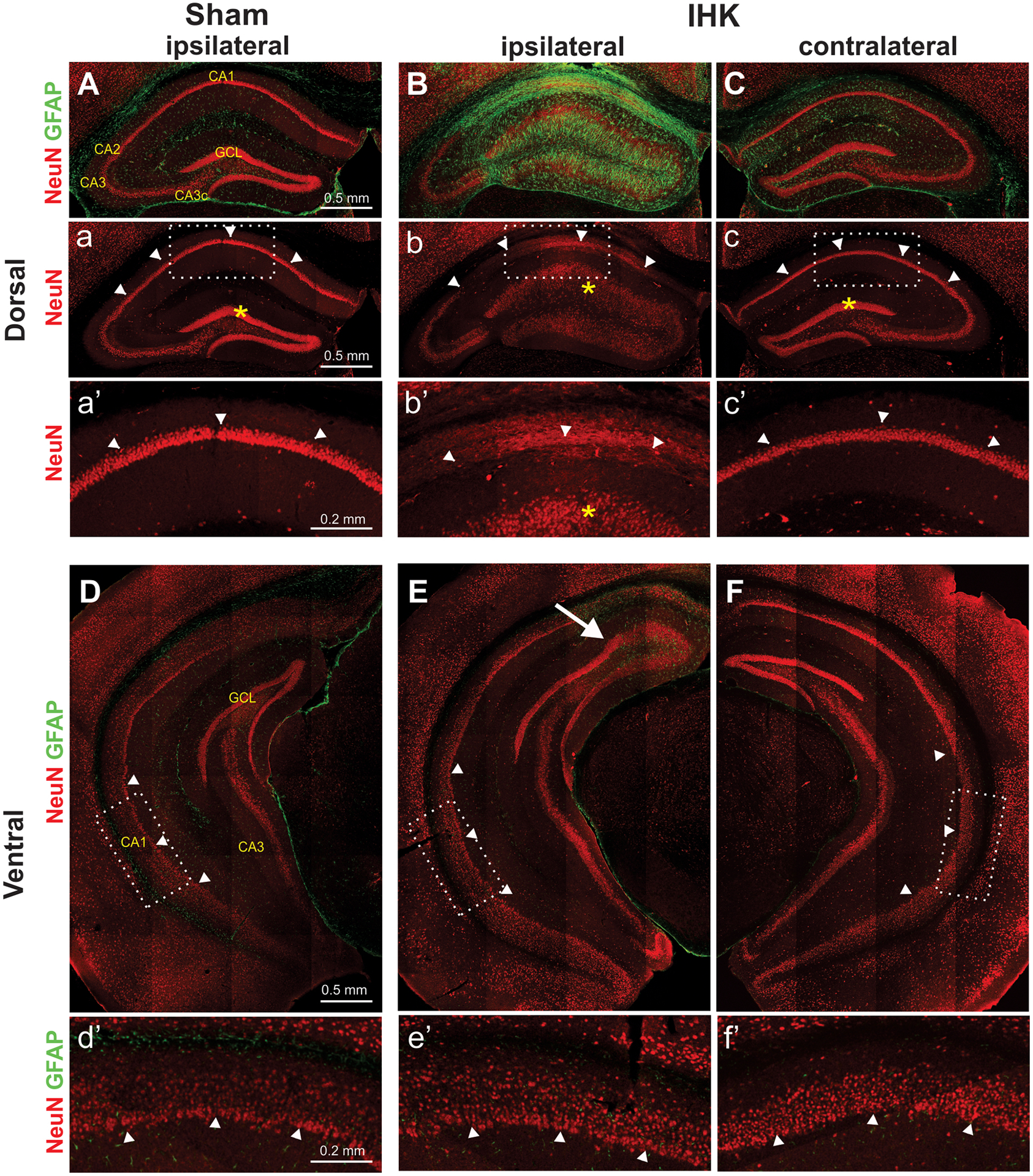
Figure 2.

Neuropathological findings in the unilateral dorsal IHK mouse model. Double immunostaining of NeuN and GFAP of hippocampi from sham control and TLE mice revealed that severe hippocampal sclerosis occurred mostly in the ipsilateral dorsal hippocampus. (A, B, C) Severe damage such as loss of CA1 and CA3c pyramidal cells, granule cell dispersion in the dentate gyrus, and GFAP-labeled gliosis was found only in the ipsilateral dorsal hippocampus of IHK mice (B, b, b’), but not in the ipsilateral hippocampus of sham controls (A, a, a’) or in the contralateral hippocampus of IHK mice (C, c, c’). These images of dorsal hippocampi were from the KA injection site (anterior-posterior: −2.0 mm from the bregma). (D, E, F) The bilateral ventral hippocampi of IHK mice appeared to be well preserved without significant sclerotic damages. There was no obvious loss of CA1 PCs in the ipsilateral ventral hippocampus from IHK mice. The images of the ventral hippocampus were taken approximately 3.2 mm posterior to bregma. The rectangular areas in “a” to “c” and “D” to “F” were enlarged and displayed in a’ to c’ and d’ to f’, respectively. Arrowheads indicate pyramidal cell layers of the CA1 region, and asterisks indicate the granule cell layer (GCL) of the dentate gyrus. The arrow in E indicates the border of a sclerotic lesion expanded from the KA injection site.
