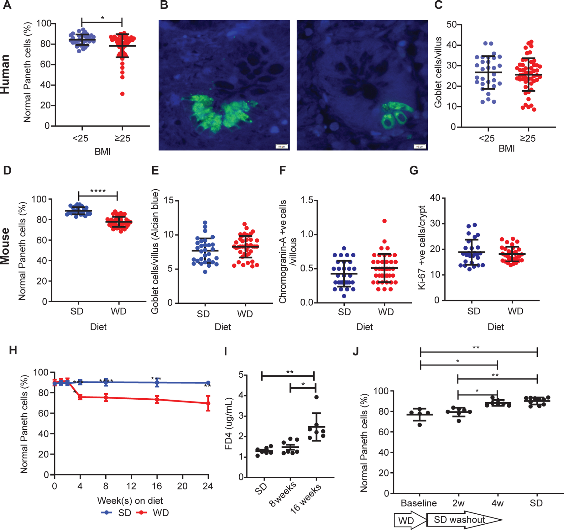
Figure 1. Obesity is associated with Paneth cell defects in humans and mice.

(A) In non-IBD patients, those with BMI≥25 (n=57) showed reduced percentage of normal Paneth cells compared to those with BMI<25 (n=34) (P=0.0129). Representative images of Paneth cells from patients with BMI<25 and BMI≥25 stained with HD5 immunofluorescence (green) are shown in (B). Scale bars: 10μm. (C) There was no difference in goblet cell density (P=0.6256) between the 2 groups. Compared to standard diet (SD)-fed mice (n=30), mice fed with Western diet (WD; n=41) for 8 weeks also resulted in (D) reduced percentage of normal Paneth cells (P<0.0001), and no significant changes in (E) goblet cell density/villus (P=0.1767), (F) neuroendocrine cells/villus (P=0.1193), or (G) crypt base proliferation (P=0.8982). (H) A time course study showed that 4 weeks of WD was sufficient to trigger Paneth cell defects (P<0.0001), whereas (I) WD only induced significant permeability change at 16 weeks (P=0.0022). (J) A 4-week washout period was sufficient to restore the percentage of normal Paneth cells (P=0.0198 compared to baseline). (H): n=3~10/group. (I): n=7/group. (J): baseline: n=5; 2 and 4wk: n=7; SD control: n=10. (A, C-G): Statistical analysis was performed by Mann-Whitney test. (H): Statistical analysis was performed by two-way ANOVA. (I, J): Statistical analysis was performed by Kruskal-Wallis test. *: P<0.05; **: P<0.01; ****: P<0.0001. Error bars represent standard deviations.
