
Figure 6. Type I IFN mediates WD-associated Paneth cell defects.
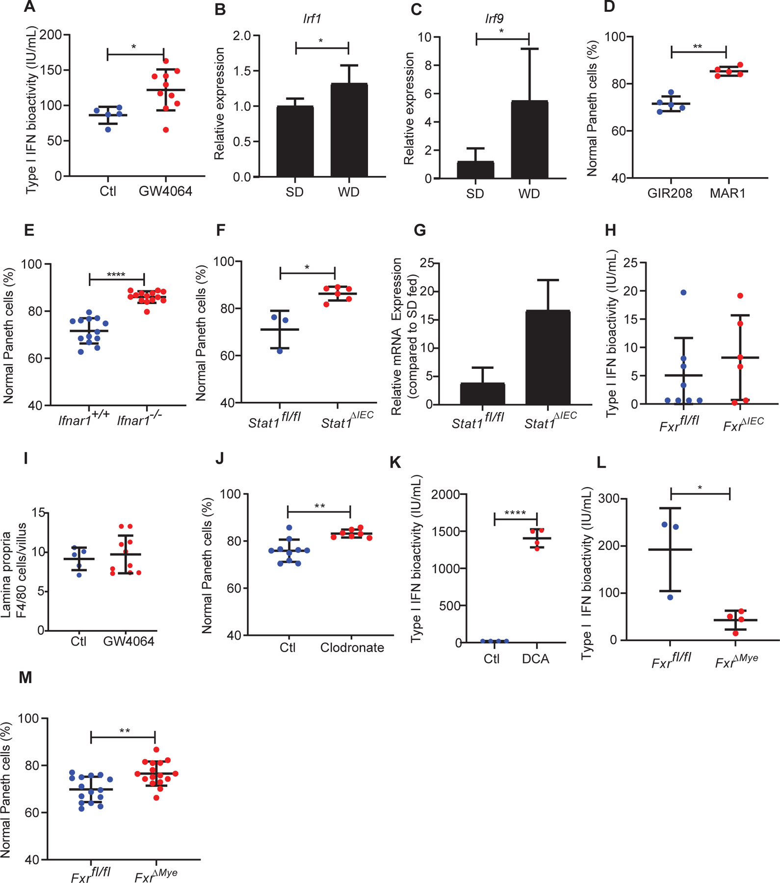
(A) Administration of GW4064 in WT, SD-fed mice showed induction of type I IFN (Ctl: n=5; GW4064: n=10; P=0.0193). This was associated with enhanced expression of type I IFN associated genes (B) Irf1 and (C) Irf9 in the crypt base compartment. (D) Treatment with anti-Ifnar1 (MAR1–5A3) but not isotype control antibody (GIR208) prevented WD-induced Paneth cell defects (n=5/group; P=0.0079). (E) Ifnar−/− mice were protected from WD-associated Paneth cell defects (Ifnar+/+: n=13, Ifnar−/−: n=14; P<0.0001). (F) Stat1ΔIEC mice fed with WD did not develop Paneth cell defects (P=0.0238). Total n: Stat1fl/fl: n=3; Stat1ΔIEC: n=6. (G) Stat1ΔIEC mice fed with WD maintained high Fgf15 expression compared to the Stat1fl/fl mice (P=0.1). (H) FxrΔIEC mice fed with WD did not show abrogation of type I IFN induction (P=0.6523). Total n: Fxrfl/fl: n=8; FxrΔIEC: n=6. (I) GW-4064-treated mice did not show increased lamina propria F4/80+ cells (P=0.6560). (J) WD-fed WT mice treated with clodronate were protected from Paneth cell defects (P=0.0031). Total n: Ct: n=10; clodronate: n=6. (K) DCA treatment induced type I IFN activity in macrophages in vitro (P<0.0001). n=4/group. (L) FxrΔMye mice fed with WD showed abrogation of type I IFN induction (P=0.0193). Total n: Fxrfl/fl: n=3; FxrΔMye: n=4. (M) FxrΔMye mice were protected from WD-mediated Paneth cell defects (P=0.0033). Total n: Fxrfl/fl: n=15; FxrΔMye: n=16. Statistical analysis for all panel was performed by Mann-Whitney test. *: P<0.05; **: P<0.01, ****: P<0.0001. Error bars represent standard deviations.