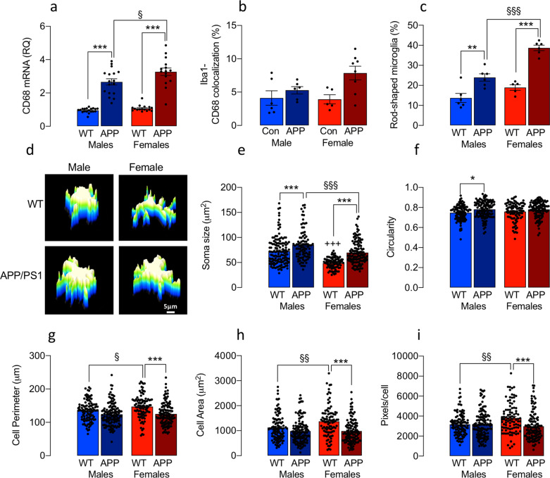
Fig. 2. Evidence of sex-related changes in microglial morphology in APP/PS1 and WT mice.
a–c CD68 mRNA (a), the co-localisation of Iba1+ CD68+ pixels (b) and the proportion of rod-shaped microglia (c) were increased in the hippocampus (and cortex, see Supplementary Fig. 2) of APP/PS1, compared with WT, mice (***p < 0.001) and a further increase was observed in female, compared with male, APP/PS1 mice (§p < 0.05; §§§p < 0.001). d 3D surface plot reconstructions show that male cells adopt an amoeboid morphology (scale bar = 5 μm). e A genotype-related increase in soma size was evident in male and female mice (***p < 0.001; d) but soma size was reduced in female WT and APP/PS1 mice compared with male counterparts (+++p < 0.001; §§§p < 0.001, respectively). f A significant main effect of genotype in circularity (p < 0.001) was observed and the mean value was significantly increased in male APP/PS1, compared with WT, mice (*p < 0.05). g–i Cell perimeter, area and pixels/cell were increased WT male, compared with the female mice (+p < 0.05; ++p < 0.01). Genotype-related decreases were observed in female mice (***p < 0.001) and changes in cell complexity were identified mainly in microglia from female mice (Supplementary Fig. 2). Data, expressed as means ± SEM (n = 5 or 6 mice/group with analysis of between 96 and 128 cells), were analysed by 2-way ANOVA and Tukey’s post hoc multiple comparison test. In the case of CD68 mRNA, data were retrospectively calculated from several previous experiments and assessed by sex (n = 15) (APP female; 16 WT female; 17 APP male; 19 WT male).

