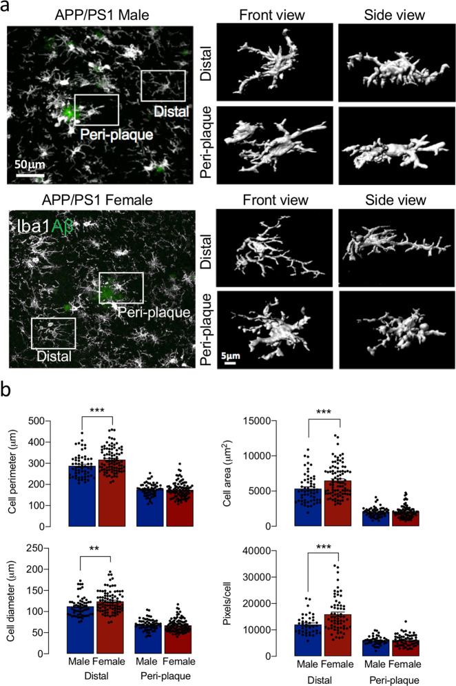
Fig. 3. Evidence of sex-related differences in morphology of microglia distal from plaques.
a 3D reconstructions showed differences in morphology in peri-plaque and distal microglia (scale bars = 50 and 5 μm in the main image and high-magnification image, respectively). b Cell perimeter, area, diameter (59–89 cells analysed) and pixels/cell (42–63 cells analysed) were markedly reduced in peri-plaque compared with distal microglia and these measures were increased in distal microglia from female, compared with male, mice (§§§p < 0.001). Data, expressed as means ± SEM (n = 5 or 6 mice/group), were analysed by 2-way ANOVA and Tukey’s post hoc multiple comparison test.

