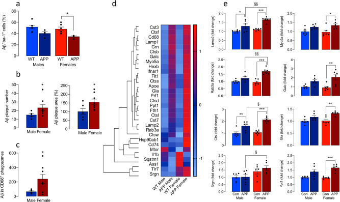
Fig. 6. Differential effect of sex on microglial function.
a Aβ uptake into isolated microglia from female APP/PS1 mice was significantly reduced compared with microglia from WT mice (*p < 0.05; n = 7, 4, 6, 5 for male WT and APP/PS1 and male WT and APP/PS1 mice, respectively). b ThioS-stained Aβ plaque number and area were significantly increased in hippocampal sections from female, compared with male, APP/PS1 mice (*p < 0.05; n = 5 and 8 for male and female mice, respectively). c Phagolysosomal loading with Aβ was significantly greater in microglia from female APP/PS1 mice compared with males (*p < 0.05; n = 6 and 8 for male and female mice, respectively). d, e NanoString analysis indicated that lysosomal genes were upregulated in microglia prepared from female APP/PS1 mice compared with the other groups (d). Analysis of the mean data, relative to values in WT males, indicated that there were significant increases in Lamp2, Myo5A, Rab3A, Ctsl, Cla, Galc, Ppt1 and Srgn in microglia from female APP/PS1 mice compared with female WT mice (*p < 0.05; **p < 0.01; ***p < 0.001) and compared with male APP/PS1 mice (§p < 0.05; §§p < 0.01; n = 6; e). Data, expressed as means ± SEM, were analysed by 2-way ANOVA and Tukey’s post hoc multiple comparison test except for b and c when the Student’s t-test for independent means was used to evaluate data.

