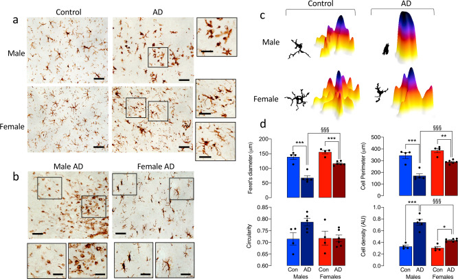
Fig. 7. Changes in microglial morphology in post-mortem parietal cortical tissue from AD patients are sex-related.
a, b Representative images of DAB-stained microglia, showing marked process retraction (a; scale bar = 100 μm; magnified image, 50 μm) and a preponderance of amoeboid microglia (b; scale bar = 200 μm; magnified image, 50 μm) in sections of parietal cortex from male AD patients compared with females, in which many rod-shaped microglia were identified. c Representative masks and 3D representations were used to analyse morphological features. d Significant disease × sex interactions in circularity, perimeter, Feret’s diameter and cell density were observed (p < 0.05). Post hoc analysis revealed significant increases in circularity and cell density and significant decreases in cell perimeter and Feret’s diameter in microglia from male AD patients compared with controls (**p < 0.01; ***p < 0.001) and also in male, compared with female, AD patients (§p < 0.05). Data, expressed as means ± SEM (n = 4 for controls or 5 for AD), were analysed by 2-way ANOVA and Tukey’s post hoc multiple comparison test.

