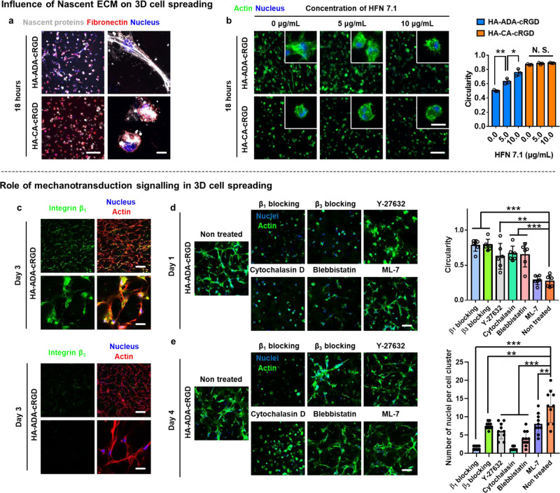
Fig. 6. The ultra-rapid cell spreading and aggregation in HA–ADA–cRGD hydrogels is regulated by cell–nascent ECM interaction, cell adhesion structures containing β1 class integrins, and actomyosin-based contractility.
a Representative images (magnifications on the right) of nascent proteins (white) and fibronectin (red) secreted by hMSCs encapsulated in HA–ADA–cRGD and HA–CA–cRGD gels within 18 h (scale bars, 200 µm (pictures on the left) and 20 μm (pictures on the right)). b Representative images (scale bars, 200 µm (main image) and 10 µm (inset)) of F-actin (green) expressed by the cells encapsulated in HA–ADA–cRGD and HA–CA–cRGD hydrogels after 18 h of treatment with different concentrations of a monoclonal antibody against the cell-adhesive domain of human fibronectin (HFN 7.1, 0, 5, 10 µg/mL, see Supplementary Fig. 19 for cell viability). Circularity of hMSCs encapsulated in HA–ADA–cRGD and HA–CA–cRGD hydrogels after 18 h of treatment with different concentrations of HFN 7.1 (0, 5, 10 µg/mL). Data are presented as mean values ± SD, n = 3 independent hydrogels, *p < 0.05, **p < 0.01 (two-tailed Student’s t-test). c Representative immunofluorescence staining against F-actin (red), nuclei (blue), and β1 class integrins or β3 class integrins (green) in hMSCs cultured in highly dynamic HA–ADA–cRGD hydrogels for 3 days (images on the top: scale bar = 200 μm. Images on the bottom: scale bar = 50 μm). d Cell spreading in the highly dynamic HA–ADA–cRGD hydrogels after 1 day of culture with or without treatment with integrin-blocking antibodies, a myosin inhibitor (blebbistatin), a myosin light chain kinase inhibitor (ML-7), a ROCK inhibitor (Y-27632), or an inhibitor of actin polymerization (Cytochalasin D). Scale bar = 100 μm. The circularity of the hMSCs encapsulated within the hydrogels treated with different inhibitors. (The average circularity value is calculated according to C = 4πA/P2, where A is the area occupied by the cell and P is the perimeter of the cell). Data are presented as mean values ± SD, n = 10 cells per group from two independent hydrogels; **p < 0.01, ***p < 0.001 (two-tailed Student’s t-test). e Multi-cell assembly in the highly dynamic HA–ADA–cRGD hydrogels after 4 days of culture with or without treatment with blocking antibodies and inhibitors. Scale bar = 100 μm. The quantification of the multicellularity of the cell clusters within the hydrogels treated with different inhibitors. Data are presented as mean values ± SD, n = 10 cell clusters per group from two independent hydrogels; **p < 0.01, ***p < 0.001 (two-tailed Student’s t-test).

