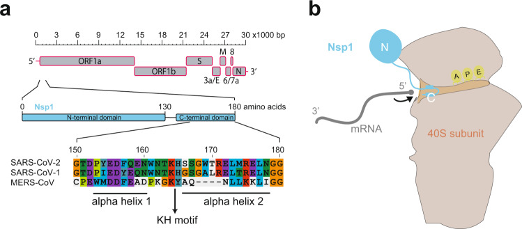
Fig. 1. Nsp1 interaction with the ribosome.
a Schematic of SARS-CoV-2 genome organisation with the whole genome depicted at the top, Nsp1 coding sequence in the middle, and a sequence alignment of Nsp1 C-terminal domain of SARS-CoV-2, SARS-CoV-1, and MERS-CoV in the lower part of the panel. The two alpha helices and the KH motif are marked by bars and an arrow, respectively. Colour coding of amino acids corresponds to default settings of the ClustalX alignment tool. b Cartoon depicting the interaction between Nsp1 and the 40S ribosomal subunit, as revealed by the structural data. The C-terminal helices anchor Nsp1 in the mRNA entry channel, thereby blocking access for host transcripts (schematically represented in grey). The globular N-terminus is not sufficiently resolved in the structures to be able to assign a clear position and function.

