Figure 3.
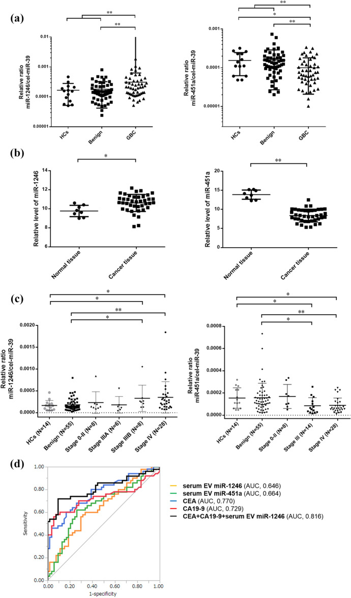
The expression levels of miR-1246 and miR-451a in serum EVs and tissues, and the potential for the application of these miRNAs as diagnostic biomarkers for gallbladder cancer. (a) The miR-1246 expression levels in serum EVs in the GBC were significantly higher in comparison to the Benign and the HCs (P = 0.005), while the miR-451a expression levels in the GBC were significantly lower in comparison to the Benign and HCs (P = 0.001). (b) The miR-1246 was significantly upregulated in gallbladder cancer tissues in comparison to normal tissue (FC = 1.79, P = 0.029), and the miR-451a was significantly downregulated in the cancer tissues in comparison to normal tissue (FC = 0.022, P < 0.001) in GSE 104165 from GEO dataset. (c) The expression levels of miR-1246 were significantly higher in GBC patients with stage IIIB–IV in comparison to the Benign and HCs, but not in patients with stage 0–IIIA. Similarly, the miR-451a expression levels was significantly lower in GBC patients with stage III–IV, but not those with stage 0–II, in comparison to the Benign and the HCs. (d) The receiver-operating characteristic (ROC) curve analysis of miR-1246, miR-451a, CEA, and CA19-9 for discriminating GBC from Benign and HCs. These curves revealed that the optimal combination was CEA, CA19-9 and miR-1246 in serum EVs (sensitivity, 72.0%; specificity, 90.8%; accuracy, 81.7%; AUC, 0.816 [95% confidence interval, 0.712–0.888]). *P < 0.05 and **P < 0.01.

