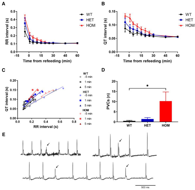
Figure 3.
Electrocardiogram telemetry during refeeding. WT (n = 8), HET (n = 7) and HOM (n = 4) mice were fasted overnight for 18 h, after which they were re-fed. (A, B) RR interval (A) and QT interval (B) dropped immediately upon refeeding. (C) RR/QT relationships were plotted and revealed a change after refeeding in HET and HOM mice, but not in WT mice. (D) During the first 30 min after refeeding, PVCs were counted, revealing an increased number of PVCs in HOM mice. (E) Representative ECG traces with PVCs in three different mice (PVCs indicated by arrowheads). Analyzed with Kruskal–Wallis test with Dunn's multiple comparisons testing. *: WT versus HOM, P < 0.05. HET, heterozygous; HOM, homozygous; PVC, premature ventricular contraction; WT, wild-type.

