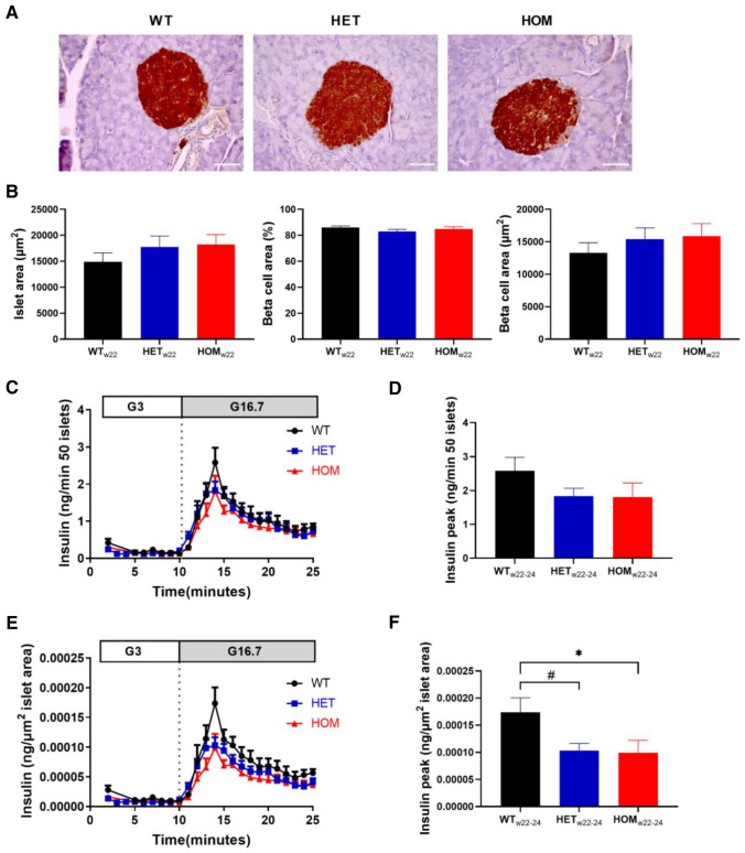
Figure 5.
Ex vivo glucose-stimulated insulin secretion and islet area in 22–24 week old mice. (A, B) Pancreatic sections of mice at 22 weeks of age were stained for insulin from WT (n = 6), HET (n = 9) and HOM (n = 7) mice, as shown in representative images (scale bar: 50 µm) (A). Islet area, the percentage of beta-cell area, and the absolute beta-cell area were measured in 5 islets per mouse and subsequently averaged per mouse. (C) Isolated islets from 22 to 24 week old WT (n = 7), HET (n = 9) and HOM (n = 8) mice were first exposed to 3 and then 16.7 M glucose. Insulin was measured in the perifusate and comparisons of peak insulin levels are depicted (D). (E) Glucose-stimulated insulin secretion was normalized to islet area measured in (B) and comparisons of peak insulin levels are depicted (F). Tested with one-way ANOVA with Dunnett’s multiple comparisons testing. *: WT versus HOM, P < 0.05; #: WT versus HET, P < 0.05. ANOVA, analysis of variance; HET, heterozygous; HOM, homozygous; WT, wild-type.

