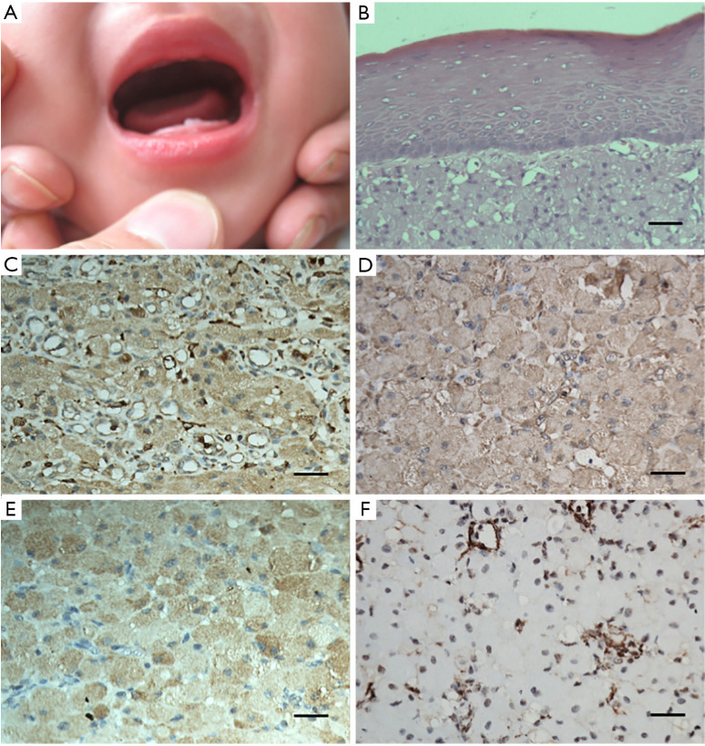
Figure 2.
The prognosis and the immunohistochemical outcomes of the reported case. (A) At the 6-month follow-up, there was no evidence of recurrence. (B) Rounded and polygonal cells with abundant granular eosinophilic cytoplasm and round or oval nuclei were observed in the lesion. Delicate connective tissue septa with small vessels were scattered throughout the granular cell lesion (H&E, ×400). (C) Positive expression of CD-68 (immunohistochemical staining, ×400). (D) Intense positive expression of vimentin (immunohistochemical staining, ×400). (E) Positive expression of NSE (immunohistochemical staining, ×400). (F) Positive expression of NK1/C3 (immunohistochemical staining, ×400). The scale bars indicate 100 µm.

