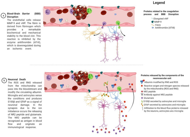
Figure 4.
Proteins derived from the proposed panel of biomarkers and their relation to the pathophysiology of stroke. After an injury, endothelial cells release MMP-9, which plays an essential role in the extracellular matrix and local proteolysis of leukocyte migration. In addition, endothelial cells release vWF in a globular form, which is then transformed into an elongated form by the activity of the ADAMTS13 enzyme. vWF promotes platelet adhesion to the damaged site by forming a molecular bridge between the subendothelial collagen matrix and the platelet-surface receptor complex GPIb-IX-V. Fibrinogen is released from the liver to the bloodstream and is cleaved by thrombin at the damaged site, resulting in fibrin formation. Fibrin is one of the main constituents of blood clots and provides remarkable biochemical and mechanical stability. The conversion of fibrinogen to fibrin by thrombin is inhibited by the enzyme antithrombin (AT-III), which is downregulated during an ischemic event. In addition, neutrophils and leukocytes arrive at the injured site and join with the previous proteins to form a solid structure that obstructs or reduces blood flow. These conditions lead to a shift in the neuron's metabolic conditions, mitochondrial dysfunction, excitotoxicity, and ion imbalance. The reactive oxygen and nitrogen species released from the mitochondria can pass into the bloodstream and modify the circulating albumin. On the other hand, microglia and astrocytes sense the conditions and produce S100β and GFAP to signal neuronal damage. In synapsis, because of ion imbalance, the release of the NR2 peptide and glutamate occurs. The NR2 peptide can be recognized as an antigen in the blood flow and induces an immunological response. Figure was drawn using Biorender.com.

