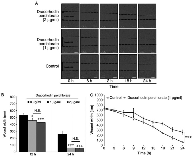
Figure 2.
Determination of cell migration in HaCaT keratinocytes. (A) Effects of dracorhodin perchlorate on wound healing of HaCaT keratinocytes. Cells (1x10⁴ cells/well) in 96-well plates were scratched and incubated with or without 1 and 2 µg/ml dracorhodin perchlorate for indicated time. (B) Cells (1x10⁴ cells/well) in a 96-well plate were scratched and treated with or without 1 and 2 µg/ml dracorhodin perchlorate for 12 and 24 h. The wound width of HaCaT keratinocytes was determined using the IncuCyte ZOOM System instrument and then quantified. (C) Time courses of the relative wound widths of HaCaT keratinocytes after wound generation. Data are presented as the mean ± standard deviation, n=3. Tukey's post hoc test after ANOVA. *P<0.05 and ***P<0.001 vs. 0 µg/ml untreated control. N.S., not significant.

