. 2021 May 3;35(3):1313–1326. doi: 10.21873/invivo.12384

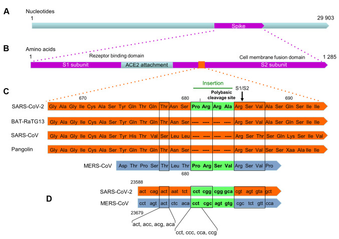
Figure 2. Sequence comparison of the spike protein of SARS-CoV-2 with other coronaviruses whose genome is very similar to that of SARS-CoV-2 (96.2% identity to the Bat-CoV RaTG13 strain isolated from the horseshoe bat; 79.5% identity to SARS-CoV and 91.0% identity to the virus isolated from the pangolin (27). (A) Genome of SARS-CoV-1. (B) S-protein. After binding to the ACE2 receptor, the spike protein is split into the S1 and S2 subunits. The S1 subunit mediates the binding to ACE2, the S2 subunit the efficient entry into the cell by membrane fusion. It is believed that after binding of the S protein to the ACE2 receptor, two (or even three) cleavage events are necessary for efficient entry into the cell. One is catalyzed by the protease TMPRSS2, the second by furin. (C) The furin cleavage site at the end of the Arg-Arg-Ala-Arg tetrapeptide is marked with an arrow (S1/S2). It is supposed that this cleavage occurs first, followed by cleavage of the S2' site (not shown) through TMPRSS2 (46). The nucleotide sequence that codes for the polybasic cleavage site (see text) rests on an insert that is unique for SARS-CoV-2. A similar insertion can be found in MERS-CoV. The amino acid sequence in the scheme is given using the 3-letter abbreviations in order to make it more understandable for nonbiochemists. Modified according to (39) and (16). (D) Sequence comparison of SARS-CoV-2 and MERS-CoV. Sequences are from NCBI database. The possible codon sequences for threonine and proline are given on the bottom of the figure.

Figure 2