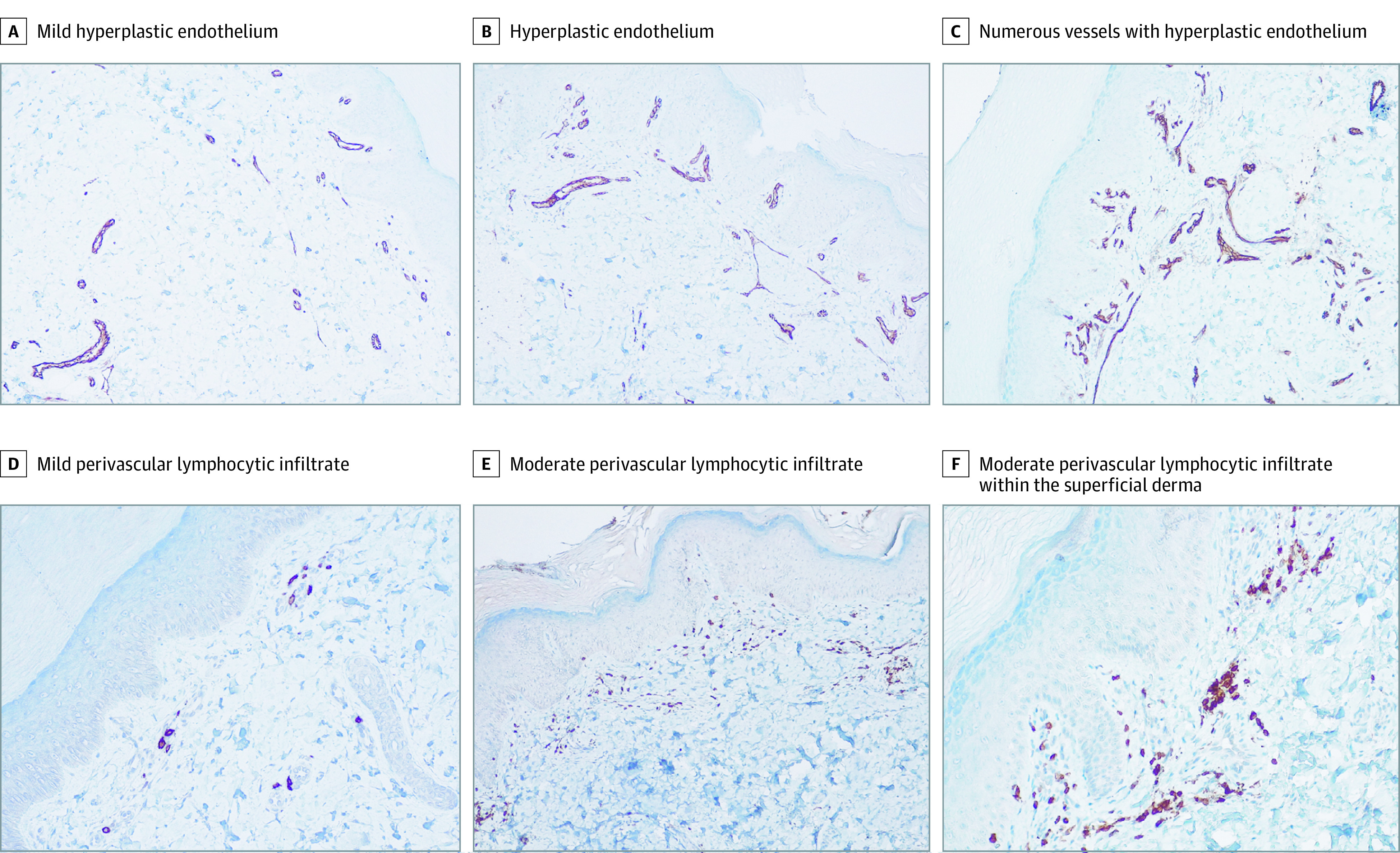
Figure 3. Microvascular Remodeling Featuring Endothelial Hyperplasia and a Mild Perivascular Lymphocytic Infiltrate.

Immunohistochemistry images from 3 representative patients (original magnification ×10). As in Figure 2, each column includes images from the same case. A-C, Endothelial cells are highlighted by anti-CD31 staining. D-F, Lymphocytes are highlighted by anti-CD3 staining.
