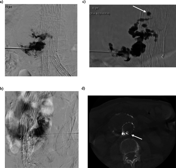
Fig. 2.
a/b Prone PA and lateral digital subtraction angiogram of the abdominal aortic sac injection with iodinated contrast through a 20G access needle demonstrates filling of the aortic sac and no feeding artery. c Prone roadmap image of the aortic sac demonstrates filling of the sac with 4:1 Lipiodol/Glue mix (arrow) and penetration through the fabric into the lumen of the graft (arrow). d Axial non-contrast cone beam CT (prone) of the abdominal aorta demonstrates non-target embolisation in the body of the stent graft. (arrow)

