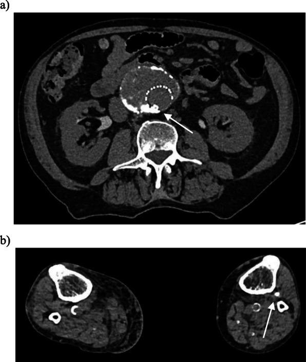
Fig. 3.

Axial non-contrast CT images of the abdomen and lower limbs performed 1 h after embolisation. Note: contrast excretion is seen from the kidneys due to contrast administered at the time of the angiography procedure. a Non-target embolisation of glue is seen (white arrow) in the stent graft lumen (arrow). b Axial non-contrast CT at the level of the calf demonstrates non-target embolization within the left anterior tibial artery
