Figure 1.
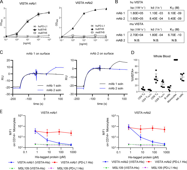
Binding and specificity of novel anti-VISTA mAbs. (A) VISTA mAb1 and VISTA mAb2 binding to plate-bound recombinant human (hu) PD-L1, huB7H5, or murine (mu) B7H5 by ELISA. (B) Binding kinetics of mAb1 and mAb2 to human and murine VISTA by SPR. No binding (N.B.) was observed for mAb2 to murine VISTA. (C) Representative Biacore sensorgram plots from simultaneous binding assay with mAb1 or mAb2 captured on the surface. Antigen binding to surface mAb is shown, followed by injection of solution mAb (soln). RU, reference units. (D) Summarized FACS staining results that quantify the percentage of VISTA+ cells in human whole blood. n = 6 independent donors. TN, naive T cells; TAg-Ex, antigen-experienced T cells. (E) FACS-based median fluorescence intensity (MFI) of isotype control, mAb1, or mAb2 binding to CD14+ PBMCs while in the presence of titrated recombinant His-tagged VISTA (VISTA-His; green triangles or blue circles) or recombinant His-tagged PD-L1 (PD-L1 His; purple triangles or red squares). A–E are representative of three independent experiments.

