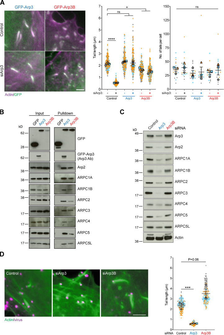
Figure 1.
Arp3 isoforms confer different cellular properties to Arp2/3. (A) Immunofluorescence images of vaccinia-induced actin tails labeled with Alexa Fluor 568 phalloidin (magenta) together with quantification of their length and number at 8 h after infection in HeLa cells stably expressing RNAi-resistant GFP-tagged Arp3 (blue) or Arp3B (red) and treated with Arp3 siRNA. (B) Immunoblot analysis of GFP-Trap pulldowns from HeLa cells lysates stably expressing GFP, GFP-Arp3, or GFP-Arp3B demonstrates that Arp3 and Arp3B associate with all Arp2/3 complex subunits. The Arp3 antibody does not detect Arp3B. (C) Immunoblot analysis of Arp2/3 complex subunits in HeLa cells treated with Arp3 and Arp3B siRNAs (Fig. S1 A shows RT-PCR analysis of the level of Arp3 and Arp3B mRNA relative to control in HeLa cells after knockdown with the individual siRNA from the siGenome pools against Arp3 and Arp3B). (D) Immunofluorescence images of virus (magenta) induced actin tails labeled with Alexa Fluor 488 phalloidin (green) together with quantification of their length in cells treated with the indicated siRNAs. All error bars represent SEM from n = 3 independent experiments in which the length of 100 tails or their number in six cells was analyzed per condition. Tukey’s multiple comparisons test was used to determine statistical significance; ****, P < 0.0001; ***, P < 0.001; *, P < 0.05. Scale bars = 5 µm.

