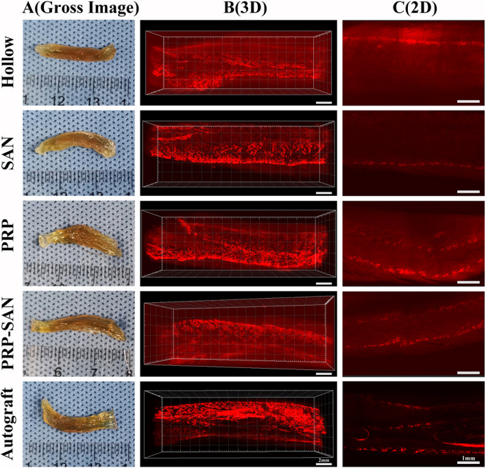
FIGURE 10.

The motor endplates (MEPs) of flexor digitorum longus at 12 weeks after surgery. (A) Representative photograph of transparency of the flexor digitorum longus after 3DISCO in each group. (B) Representative three‐dimensional distributions of MEPs in flexor digitorum longus. Scale bar =2 mm. (C) The coronal plane photograph shown in (B). Scale bar =1 mm
