
FIGURE 1.
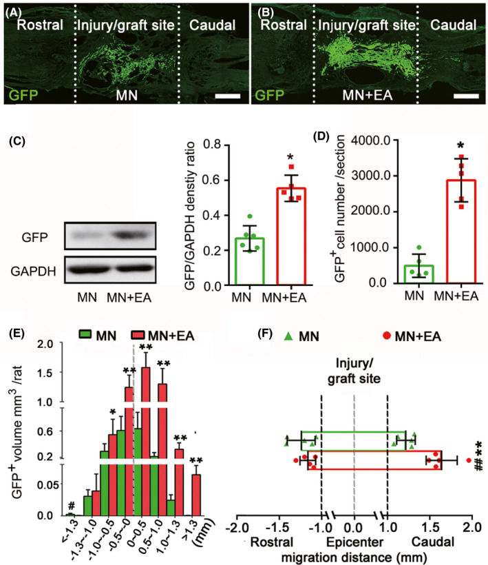
Electroacupuncture (EA) promoted the survival and migration of mesenchymal stem cell (MSC)‐derived cells in vivo. (A, B) Representative immunofluorescence images of injured spinal cord sections from the MSC‐derived neural network (MN, A) and MN+EA groups (B) at 8‐week post‐injury (wpi). Scale bars = 500 µm. Green fluorescent protein‐positive (GFP+) cells (green) are MSC‐derived cells from the grafted MN. (C) Western blot analysis of GFP expression in the injury/graft site in the MN+EA and MN groups at 2 wpi (n = 5/group). (D, E) Bar charts showing the number (D) and volume (E) of grafted GFP+ cells. Values represent the mean ± SD (n = 5/group, Student's t‐test, *p < 0.05). (F) Graphical representation showing the average maximum migration distance of GFP+ cells from the epicenter of grafted MN to the rostral and caudal sides in each group. The quantification of migration distance was performed using ImageJ. Values represent the mean ± SD (n = 5/group. *p < 0.05, **p < 0.01, and ## p < 0.01, determined by unpaired Student's t‐test). GAPDH, glyceraldehyde 3‐phosphate dehydrogenase