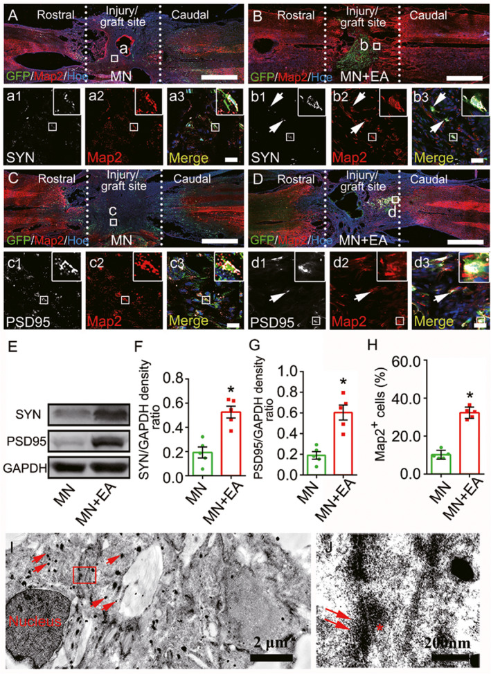
FIGURE 2.

Mesenchymal stem cell (MSC)‐derived neuron‐like cells and synapse‐like structures in the graft site of the spinal cord at 8 wpi. (A–D) Representative images triple‐labeled with anti‐green fluorescent protein (GFP, green), microtubule‐associate protein (Map2, red; a2,b2,c2, and d2) and synapsin (SYN, white; a1 and b1) or postsynaptic density protein 95 (PSD95, white; c1 and d1) in the epicenter of the injury/graft site. The merged images show the GFP+ cells that colocalize with Map2/SYN (arrows, a3 and b3) or Map2/PSD95 (arrows, c3 and d3). The cell nuclei were counterstained with Hoechst33342 (Hoe). (E–G) Western blot analysis of SYN and PSD95 expression in the injury/graft site of the spinal cord. Values represent the mean ± SD (n = 5/group, *p < 0.05). (H) Bar chart showing the percentage of GFP+ cells that differentiated into Map2+ neuron‐like cells. Values represent the mean ± SEM (n = 5/group, *p < 0.05). (I, J) IEM revealed that a GFP+ cell, labeled by silver‐enhanced nanogold particles (arrows), formed a synapse‐like structure with another GFP+ cell. The structure was characterized by the presence of some synaptic vesicles (J, arrows) of varying diameters in the “presynaptic element,” electron dense material in the “postsynaptic element” (J, asterisk), and a “synaptic cleft” between the presynaptic and postsynaptic elements. Scale bars = 1 mm in (A–D); 20 µm in (a1)–(a3), (b1)–(b3), (c1)–(c3) and (d1)–(d3)
