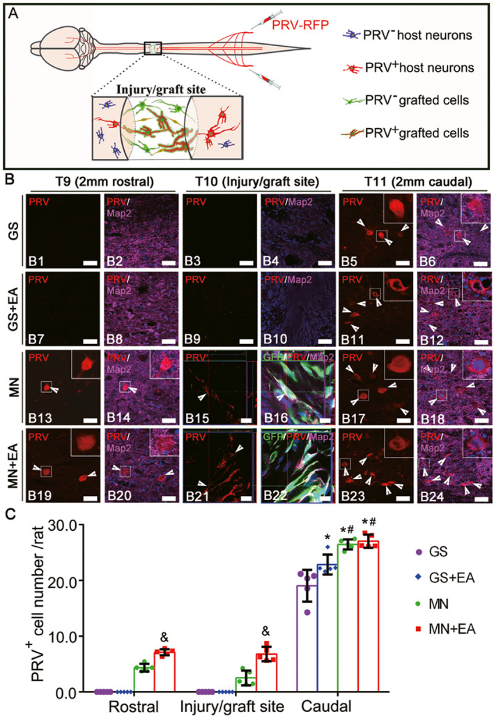
FIGURE 4.

Pseudorabies virus (PRV) retrograde transsynaptic labeling confirmed the integration of transplanted mesenchymal stem cell (MSC)‐derived neuron‐like cells into the host spinal cord neuronal circuit. (A) A schematic diagram showing that PRV that was injected into the sciatic nerve was transported from the caudal area to the rostral area through the injury/graft site of the spinal cord. (B) Representative images showing the host neurons or MSC‐derived neuron‐like cells retrogradely labeled with PRV (red, arrowheads) in the rostral and caudal regions relative to the graft tissue of spinal cord in the GS group (B1–B6), GS+EA group (B7–B12), MN group (B13–B18) and MN+EA group (B19–B24). The cell nuclei were counterstained with Hoechst33342 (Hoe). (C) Bar chart showing the number of PRV+ neurons in the T9, T10, and T11 areas of the four groups. Values represent the mean ±SD. n = 5/group. *p < 0.05, compared with the GS group, # p < 0.05, compared with the GS+EA group, and & p < 0.05, compared with the MN group by one‐way ANOVA with LSD‐t. Green fluorescent protein (GFP, green), PRV (red), microtubule‐associated protein (Map2, white), and Hoe (blue). Scale bars =50 µm in (B1)–(B14), (B17)–(B20), (B23), and (B24); 10 µm in (B15) and (B16), (B21), and (B22). GS: gelatin sponge scaffold with no cells; GS+EA: GS combined electroacupuncture; MN: MSC‐derived neural network; MN+EA: MN combined with electroacupuncture
