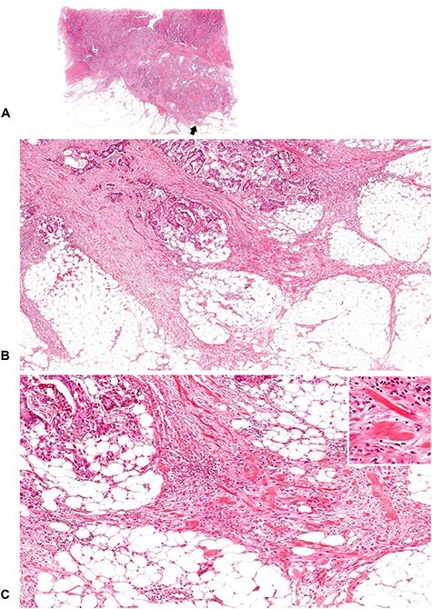
Figure 10.

A tumor with intermediate DR (2). Keloid-like collagen bundles appear at the leading edge of the desmoplastic stroma. (B; ×4 objective) and (C; ×10 objective) are magnifications of the area of the desmoplastic front as indicated by the arrow in (A) (whole slide image). Insert in (C), ×20 objective. All, H&E staining.
