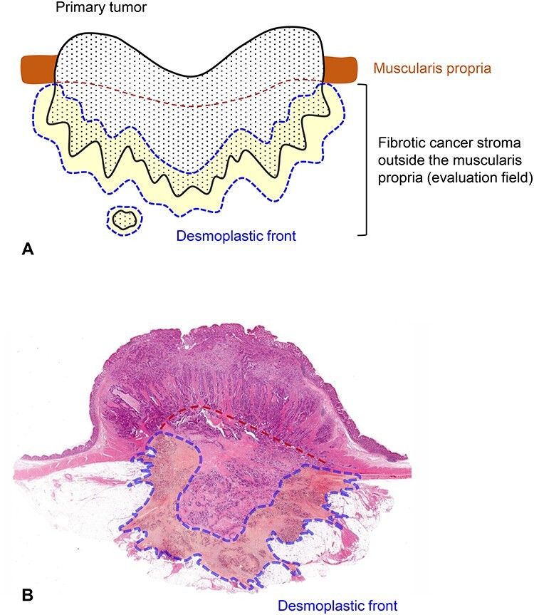
Figure 2.

Field in the primary tumor to be evaluated for the DR classification. Fibrotic cancer stroma outside the muscularis propria is evaluated on pathological slides prepared in routine practice. Cancer stroma in the submucosa or those in the muscularis propria are excluded from the evaluation. Fibrotic stroma along the leading edge of the primary tumors (desmoplastic front) is the area subject to intensive evaluation because both myxoid stroma and keloid-like hyalinized collagen mostly appear in this lesion. When a tumor nodule exists in the fatty tissue attached to the primary tumor, the fibrotic stroma of the lesion should be evaluated entirely for the DR classification. (A) Schema and (B) a whole slide image [Hematoxylin–eosin (H&E) stained] of the primary tumor. Area circled with blue dotted line, desmoplastic front; brown dotted line, estimated lower border of the muscularis propria.
