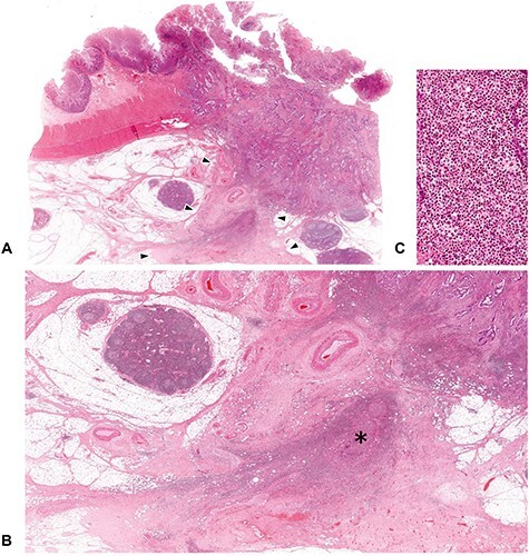
Figure 3.

Fibrotic area to be excluded from the DR classification evaluation. The DR pattern is evaluated at fibrotic stroma exclusively induced by tumor–stroma interaction. Acute inflammation such as microscopic abscess with intratumoral perforation can evoke a dense fibrosis, but this area should be distinguished from the desmoplastic reaction to be assessed for the DR classification. In figure (A), dense fibrotic tissue exists beyond the desmoplastic front (area surrounded by the triangle symbols), in which a remnant of microscopic abscess (shown with an asterisk) is observed, thereby indicates this fibrosis was induced by the external pathogens, rather than tumor–stroma interaction. (B; ×1.5 objective) is photograph magnifying the part of the extramural area indicated by the triangles in (A; whole slide image) and (C; ×40 objective) is that magnifying that part of asterisk. All, H&E staining.
