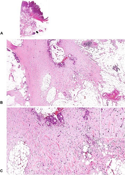
Figure 7.

A tumor with immature DR (2). An amorphous, vacuolated, amphophilic myxoid material extends to the desmoplastic front. Note that although the tumor cells produce abundant extracellular mucin, myxoid stroma can be differentiated from the mucin pool in the stroma. The mucin leakage is devoid of desmoplastic reaction, whereas the myxoid stroma is intermingled with other desmoplastic components such as fibroblasts, collagen fibers, inflammatory cells and endothelial cells. (B; ×4 objective) and (C; ×10 objective) are magnified photographs of the portion of the desmoplastic front indicated by the arrow in (A; whole slide image). Insert in (C), ×20 objective. All, H&E staining.
