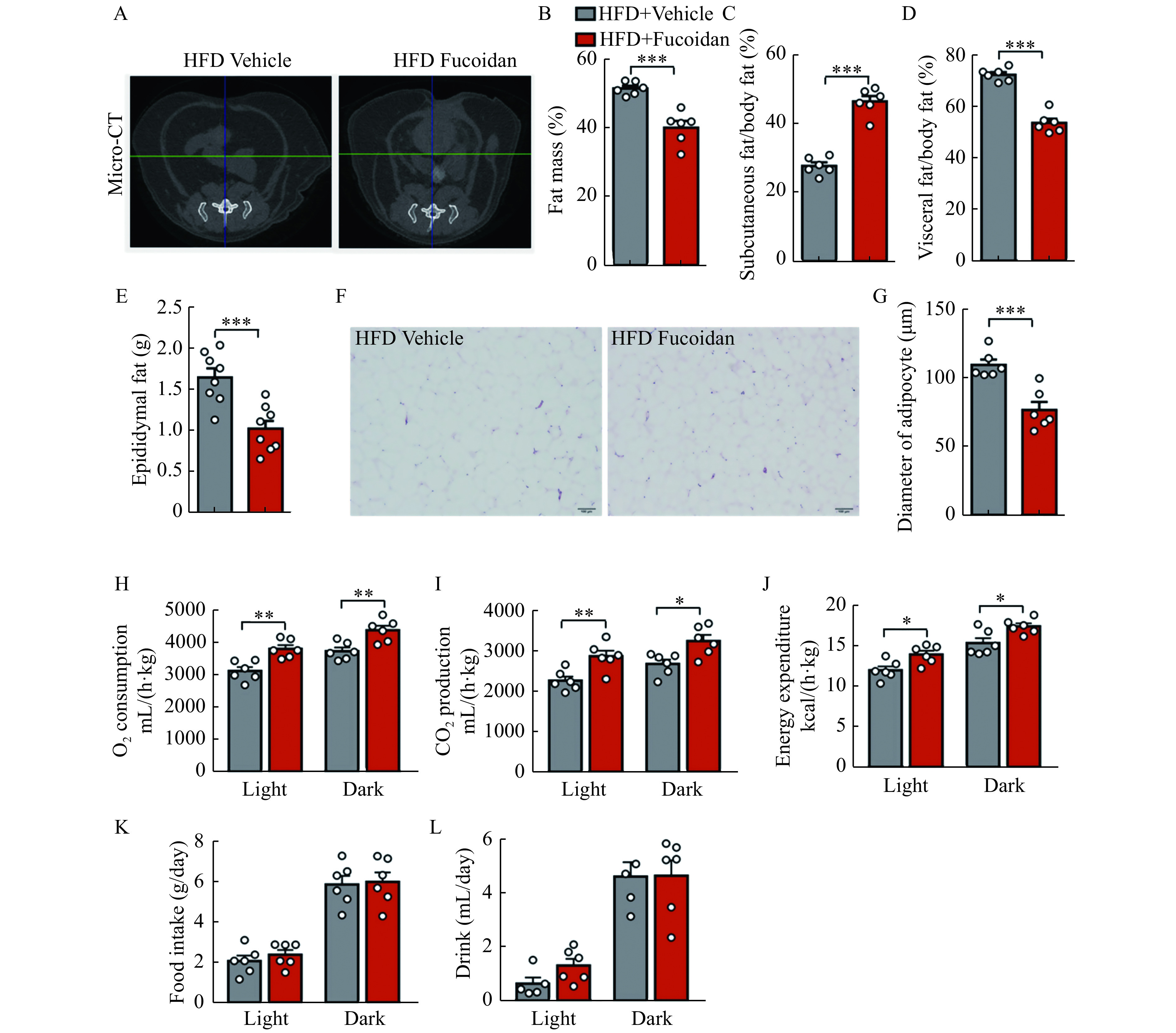
Figure 2.

Fucoidan treatment suppressed adipose hypertrophy.
Mice were administered with fucoidan after 8 weeks of HFD feeding. A: Representative micro-CT images of the abdominal fat at L6 region in fucoidan-treated mice after 24 weeks of HFD feeding. B–D: Fat mass percentage (B), subcutaneous fat percentage (C), and visceral fat percentage (D) in the fucoidan-treated mice after 24 weeks of HFD feeding were measured by micro-CT,n=6. E: Epididymal fat weight in fucoidan-treated mice after 24 weeks of HFD feeding, n=8. F: Representative images of H&E staining of epididymal fat tissues in the fucoidan-treated mice after 24 weeks of HFD feeding. Scale bars, 100 μm. G: Average diameters of adipocytes in epididymal adipose tissue of fucoidan-treated mice after 24 weeks of HFD feeding,n=6. H–L: whole body oxygen (O2) consumption (H), carbon dioxide (CO2) production (I), energy expenditure (J), food intake (K), and drink (L) in fucoidan-treated mice after 24 weeks of HFD feeding during a 24-hour period. Data are expressed as mean±SEM. Statistical analyses were performed by two-tailed unpaired t test for two-group comparisons. *P<0.05;**P<0.01;***P<0.001. HFD: a high-fat diet; micro-CT: micro-computer tomography.
