Figure 2.

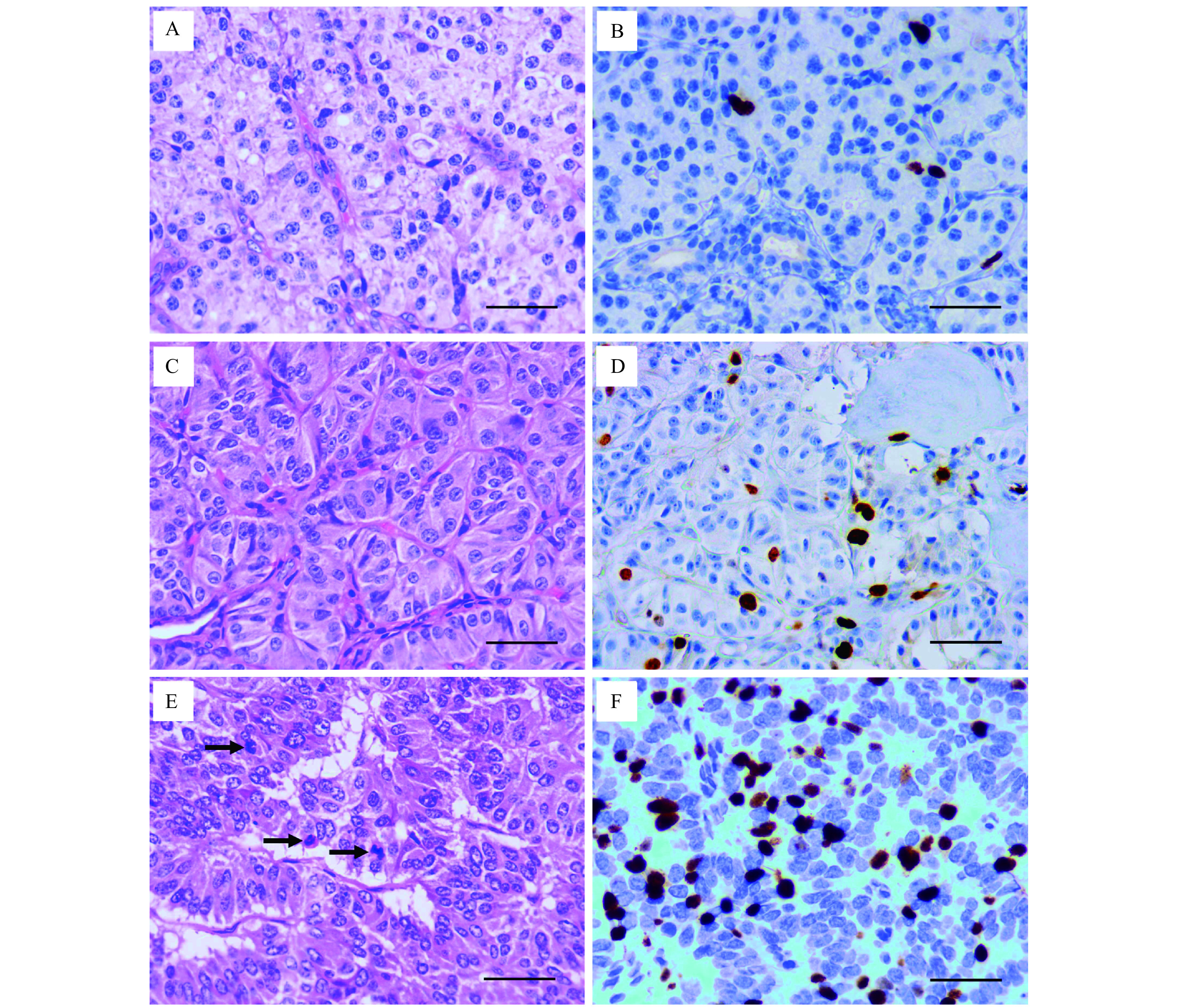
Hematoxylin-eosin and Ki-67 staining of pancreatic neuroendocrine neoplasms.
A: Mitoses <2 per 10 HPF in Hematoxylin-eosin (HE) staining of PNENs G1. B: Ki-67 index <3% in immunohistochemical (IHC) staining of PNENs G1. C: Mitoses 2–20 per 10 HPF in HE staining of PNENs G2. D: Ki-67 index 3%–20% in IHC staining of PNENs G2. E: Mitoses >20 per 10 HPF in HE staining of PNENs G3. Black arrows indicate the mitotic cells. F: Ki-67 index >20% in IHC staining of PNENs G3. Scale bar=50 μm. PNENs: pancreatic neuroendocrine neoplasms; HPF: high power field.
