
Figure 3.
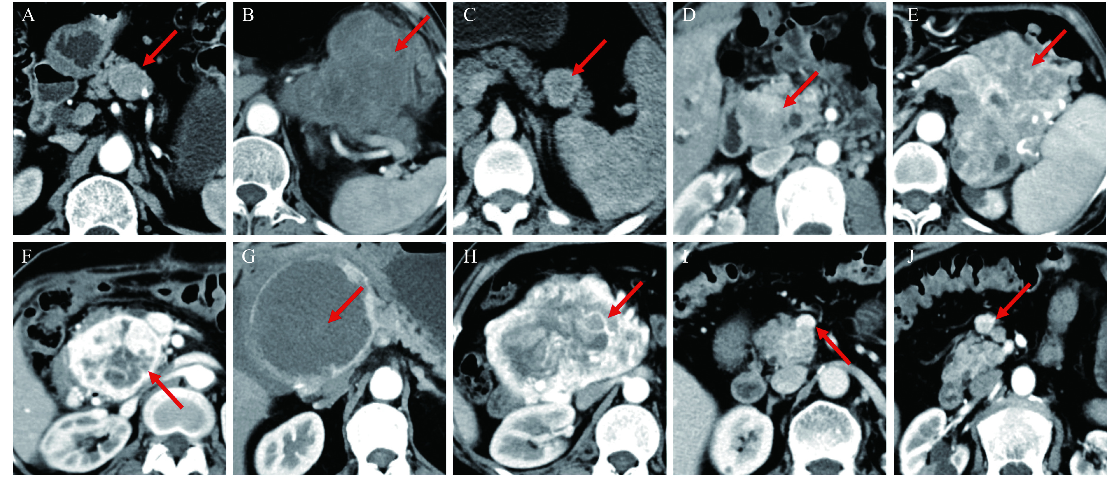
The imaging features of pancreatic neuroendocrine neoplasms on contrast-enhanced CT.
A: Regular tumor shape and iso-density in arterial phase. B: Irregular tumor shape and hypo-density in arterial phase. C: Well-defined tumor margin. D: Ill-defined tumor margin. E: Heterogeneous enhancement. F: Edge enhancement. G: Cystic or necrotic components. H: Intratumoral blood vessels. I: Vascular invasion. J: Lymph node metastases. CT: computed tomography.