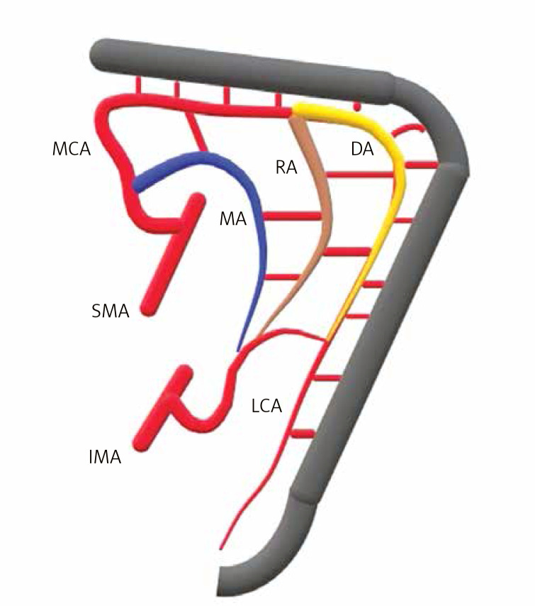
Figure 1.
Upper image represents the collateral structures at the splenic flexure level; the yellow marked structure at the lateral side is the Drummond marginal artery (DA), the brown marked structure in the middle is Riolan’s arch (RA), and the blue marked structure at the medial side is the Moskowitz artery (MA)
MCA – middle colic artery, RA – Riolan’s arch, DA – Drummond artery, MA – Moskowitz artery, SMA – superior mesenteric artery, IMA – inferior mesenteric artery, LCA – left colic artery.

