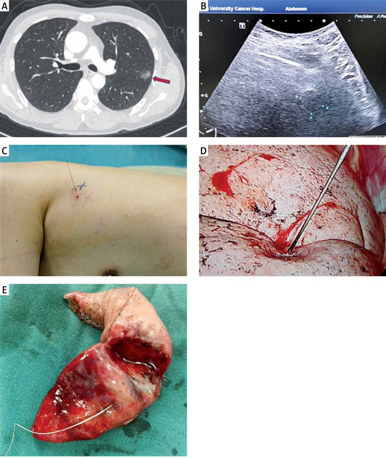
Photo 1.
A – Preoperative high-resolution CT showed mGGO of the upper left lung, with a size of 10 × 13 mm (red arrow), a shortest distance skin marker was made (blue dot). B – Percutaneous ultrasound was performed near the CT marker to detect the subpleural nodules (10 mm below visceral pleura, green dot) during the operation, and guide the hook wire to the mGGO. C – CT maker (blue dot) and ultrasonic marker (red dot) have small distance between each other and a good repeatability. D – Hook wire needle was observed under uniportal thoracoscopy, and a pulmonary wedge resection was performed at least 2 cm away from the puncture point. E – Postoperative subpleural nodules were found with naked eye, and pathological indicated infiltrating adenocarcinoma. An extended lobectomy was performed

