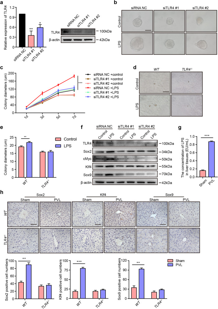
Fig. 5.
LPS promotes colony formation and pluripotent marker expression via the TLR4 pathway. a The relative expression of TLR4 at the RNA and protein levels in AML12 cells after transfection with siRNA NC, siTLR4 #1, and siTLR4 #2. b, c After transfection with siRNA NC or siTLR4 for 24 h, AML12 cells were plated and cultured with or without LPS for 7 days. Representative images of colonies (b) and the quantification of colony diameters (c). d Representative images of primary hepatocytes from WT and TLR4-/- mice cultured with or without LPS for 2 weeks. e Quantification of the colony diameters of primary hepatocytes from WT or TLR4-/- mice. f After transfection with siRNA NC or siTLR4 for 24 h, AML12 cells were cultured with LPS for 24 h and lysed to analyze the expression of TLR4, Sox2, cMyc, Klf4, and Sox9 by Western blot. g After ligation of the left portal vein branch, the right liver lobes were obtained from the sham and PVL groups. The concentrations of LPS in the right liver lobes of WT mice from the sham and PVL groups. h Upper: IHC staining of Sox2, Klf4, and Sox9 in the right liver lobes of WT and TLR4-/- mice in the sham and PVL groups. Bottom: Quantification of the Sox2-, Klf4-, and Sox9-positive cell numbers. Scale bars, 50 μm. d: days. *P < 0.05, **P < 0.01, ***P < 0.001

